PDCD1LG2
PDCD1LG2| PDCD1LG2 | |||||||||||||||||||||||||
|---|---|---|---|---|---|---|---|---|---|---|---|---|---|---|---|---|---|---|---|---|---|---|---|---|---|
| 식별자 | |||||||||||||||||||||||||
| 별칭 | PDCD1LG2, B7DC, Btdc, CD273, PD-L2, PDCD1L2, PDL2, bA574F11.2, 프로그래밍된 셀 데스 1리간드 2 | ||||||||||||||||||||||||
| 외부 ID | OMIM: 605723 MGI: 1930125 HomoloGene: 10973 GeneCard: PDCD1LG2 | ||||||||||||||||||||||||
| |||||||||||||||||||||||||
| |||||||||||||||||||||||||
| |||||||||||||||||||||||||
| |||||||||||||||||||||||||
| 직교체 | |||||||||||||||||||||||||
| 종 | 인간 | 마우스 | |||||||||||||||||||||||
| 엔트레스 | |||||||||||||||||||||||||
| 앙상블 | |||||||||||||||||||||||||
| 유니프로트 | |||||||||||||||||||||||||
| RefSeq(mRNA) | |||||||||||||||||||||||||
| RefSeq(단백질) | |||||||||||||||||||||||||
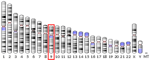
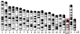
| 위치(UCSC) | Cr 9: 5.51 – 5.57Mb | Cr 19: 29.39 – 29.45Mb | |||||||||||||||||||||||
| PubMed 검색 | [3] | [4] | |||||||||||||||||||||||
| 위키다타 | |||||||||||||||||||||||||
| |||||||||||||||||||||||||
프로그래밍된 세포 데스 1 리간드 2(PD-L2, B7-DC라고도 함)는 인간에서 PDCD1LG2 유전자에 의해 암호화된 단백질이다.[5][6]PDCD1LG2도 CD273(분화 273 클러스터)으로 지정되었다.PDCD1LG2는 면역 체크포인트 수용체 리간드로, 적응성 면역 반응의 음성 조절에 역할을 한다.[5][7]PD-L2는 프로그램된 세포사멸단백질 1(PD-1)을 위한 2개의 알려진 리간드 중 하나이다.[5]
구조

PD-L2는 B7 단백질군에 속하는 세포표면수용체다.[8]면역글로불린과 같은 가변영역과 세포외 영역의 면역글로불린과 같은 상수영역, 투과영역, 세포질영역으로 구성된다.[8]PD-L2는 다른 B7 단백질과 상당한 시퀀스 호몰로학을 공유하지만 CD28/CTLA4, 즉 SQDXXXELY 또는 XXXXXRT에 대한 putive binding sequence를 포함하지 않는다.[9][9]
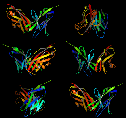
무두질 PD-1에 바인딩된 무두질 PD-L2의 결정구조가 결정되었다.[10]hPD-L2/mutant hPD-1 단지의 [11]구조뿐 아니라
표현
프로필
PD-L2는 주로 덴드리트 세포(DC)와 대식세포(대식세포)를 포함한 전문 항원 제시 세포에 표현된다.[12]다른 이들은 특정 T 도우미 세포 서브셋과 세포독성 T 세포에서 PD-L2 표현을 보여주었다.[13][14]PD-L2 단백질은 GI트랙트 조직, 골격근, 편도선, 췌장을 포함한 많은 건강한 조직에서 널리 표현된다.[15]또한 PD-L2는 3중 음성 유방암과 위암에서는 보통에서 높은 발현을 하고 신장세포암에서는 낮은 발현을 한다.[16]PD-L2 mRNA는 널리 표현되며 특정 조직에서 농축되지 않는다.[15]
규정
인터루킨-4(IL-4)와 그래눌로시세포군 군집 자극 인자(GMCSF)는 모두 시험관내 DC에서 PD-L2 발현을 상향 조절한다.[12]IFN-α, IFN-β, IFN-극복은 PD-L2 표현식의 적절한 상향 조정을 유도한다.[12]
함수
PD-L2는 11.3nM의 분해 상수 K로d 수용체 PD-1에 바인딩된다.[17] PD-1에 바인딩하면 TCR/BCR 매개 면역세포 활성화를[12] 억제하는 경로를 활성화할 수 있다(자세한 내용은 PD-1 신호 참조).PD-L2는 면역 내성과 자기 면역성에 중요한 역할을 한다.[18]PD-L1과 PD-L2 모두 T세포 증식과 염증성 사이토카인 생산을 억제할 수 있다.[17]PD-L2 차단은 실험용 자가면역 뇌근막염을 악화시키는 것으로 나타났다.[18]PD-L1과 달리 PD-L2는 면역체계를 활성화하는 것으로 나타났다.PD-L2는 뮤린 덴드리트 셀에서 IL-12 생산을 촉발하여 T 셀을 활성화시킨다.[17]다른 이들은 PD-L2 Ig를 이용한 치료가 T 도우미 세포 증식을 초래했다는 것을 보여주었다.[18]
임상적 유의성
특정 암에 대한 면역 반응에서 PD-L2, PD-L1, PD-1 표현이 중요하다.적응성 면역체계를 억제하는 역할 때문에 PD-1과 PD-L1을 차단하기 위한 노력이 이루어져 FDA가 승인한 억제제(펨브로리주맙, 니볼루맙, 아톨리주맙 참조)가 있다.2019년 현재 PD-L2에 대한 FDA 승인 억제제는 아직 없다.[19]
암 진행과 면역-투명 미세환경 규제에서 PD-L2의 직접적인 역할은 PD-L1의 역할만큼 잘 연구되지 않는다.[16]마우스 세포 배양에서는 종양 세포에 대한 PD-L2 발현이 세포독성 T 세포 매개 면역 반응을 억제했다.[20]
간접적으로 PD-L2는 바이오마커 또는 예측지표로 효용성을 가질 수 있다.PD-L2 표현은 PD-L1 표현과 독립적으로 펨브로리주맙을 이용한 PD-1 봉쇄에 대한 대응을 예측하는 것으로 나타났다.[16]그러나 PD-L2는 암의 결과를 putpically 예측하지 않으며, 일부 연구에서는 음성 예후를[21][22][23] 예측하고 다른 연구에서는 양성 예후를 예측하고 있다.[24]
참조
- ^ a b c GRCh38: 앙상블 릴리스 89: ENSG00000197646 - 앙상블, 2017년 5월
- ^ a b c GRCm38: 앙상블 릴리스 89: ENSMUSG000016498 - 앙상블, 2017년 5월
- ^ "Human PubMed Reference:". National Center for Biotechnology Information, U.S. National Library of Medicine.
- ^ "Mouse PubMed Reference:". National Center for Biotechnology Information, U.S. National Library of Medicine.
- ^ a b c Latchman Y, Wood CR, Chernova T, Chaudhary D, Borde M, Chernova I, et al. (March 2001). "PD-L2 is a second ligand for PD-1 and inhibits T cell activation". Nature Immunology. 2 (3): 261–8. doi:10.1038/85330. PMID 11224527. S2CID 27659586.
- ^ "Entrez Gene: PDCD1LG2 programmed cell death 1 ligand 2".
- ^ McDermott DF, Atkins MB (October 2013). "PD-1 as a potential target in cancer therapy". Cancer Medicine. 2 (5): 662–73. doi:10.1002/cam4.106. PMC 3892798. PMID 24403232.
- ^ a b Chen L (May 2004). "Co-inhibitory molecules of the B7-CD28 family in the control of T-cell immunity". Nature Reviews. Immunology. 4 (5): 336–47. doi:10.1038/nri1349. PMID 15122199. S2CID 33548210.
- ^ a b Tseng SY, Otsuji M, Gorski K, Huang X, Slansky JE, Pai SI, et al. (April 2001). "B7-DC, a new dendritic cell molecule with potent costimulatory properties for T cells". The Journal of Experimental Medicine. 193 (7): 839–46. doi:10.1084/jem.193.7.839. PMC 2193370. PMID 11283156.
- ^ Lázár-Molnár E, Yan Q, Cao E, Ramagopal U, Nathenson SG, Almo SC (July 2008). "Crystal structure of the complex between programmed death-1 (PD-1) and its ligand PD-L2". Proceedings of the National Academy of Sciences of the United States of America. 105 (30): 10483–8. doi:10.1073/pnas.0804453105. PMC 2492495. PMID 18641123.
- ^ Tang S, Kim PS (December 2019). "A high-affinity human PD-1/PD-L2 complex informs avenues for small-molecule immune checkpoint drug discovery". Proceedings of the National Academy of Sciences of the United States of America. 116 (49): 24500–24506. doi:10.1073/pnas.1916916116. PMC 6900541. PMID 31727844.
- ^ a b c d Sharpe AH, Wherry EJ, Ahmed R, Freeman GJ (March 2007). "The function of programmed cell death 1 and its ligands in regulating autoimmunity and infection". Nature Immunology. 8 (3): 239–45. doi:10.1038/ni1443. PMID 17304234. S2CID 8749576.
- ^ Messal N, Serriari NE, Pastor S, Nunès JA, Olive D (September 2011). "PD-L2 is expressed on activated human T cells and regulates their function" (PDF). Molecular Immunology. 48 (15–16): 2214–9. doi:10.1016/j.molimm.2011.06.436. PMID 21752471.
- ^ Lesterhuis WJ, Steer H, Lake RA (October 2011). "PD-L2 is predominantly expressed by Th2 cells". Molecular Immunology. 49 (1–2): 1–3. doi:10.1016/j.molimm.2011.09.014. PMID 22000002.
- ^ a b "Tissue expression of PDCD1LG2". The Human Protein Atlas. Retrieved 2020-03-05.
- ^ a b c Yearley JH, Gibson C, Yu N, Moon C, Murphy E, Juco J, et al. (June 2017). "PD-L2 Expression in Human Tumors: Relevance to Anti-PD-1 Therapy in Cancer". Clinical Cancer Research. 23 (12): 3158–3167. doi:10.1158/1078-0432.CCR-16-1761. PMID 28619999.
- ^ a b c Ghiotto M, Gauthier L, Serriari N, Pastor S, Truneh A, Nunès JA, Olive D (August 2010). "PD-L1 and PD-L2 differ in their molecular mechanisms of interaction with PD-1". International Immunology. 22 (8): 651–60. doi:10.1093/intimm/dxq049. PMC 3168865. PMID 20587542.
- ^ a b c Zhang Y, Chung Y, Bishop C, Daugherty B, Chute H, Holst P, et al. (August 2006). "Regulation of T cell activation and tolerance by PDL2". Proceedings of the National Academy of Sciences of the United States of America. 103 (31): 11695–700. Bibcode:2006PNAS..10311695Z. doi:10.1073/pnas.0601347103. PMC 1544232. PMID 16864790.
- ^ "Search of: PDCD1LG2 - List Results - ClinicalTrials.gov". clinicaltrials.gov. Retrieved 2020-03-04.
- ^ Tanegashima T, Togashi Y, Azuma K, Kawahara A, Ideguchi K, Sugiyama D, et al. (August 2019). "Immune Suppression by PD-L2 against Spontaneous and Treatment-Related Antitumor Immunity". Clinical Cancer Research. 25 (15): 4808–4819. doi:10.1158/1078-0432.CCR-18-3991. PMID 31076547.
- ^ Wang ZL, Li GZ, Wang QW, Bao ZS, Wang Z, Zhang CB, Jiang T (2019). "PD-L2 expression is correlated with the molecular and clinical features of glioma, and acts as an unfavorable prognostic factor". Oncoimmunology. 8 (2): e1541535. doi:10.1080/2162402X.2018.1541535. PMC 6343813. PMID 30713802.
- ^ Yang H, Zhou X, Sun L, Mao Y (2019). "Correlation Between PD-L2 Expression and Clinical Outcome in Solid Cancer Patients: A Meta-Analysis". Frontiers in Oncology. 9: 47. doi:10.3389/fonc.2019.00047. PMC 6413700. PMID 30891423.
- ^ Tobin JW, Keane C, Gunawardana J, Mollee P, Birch S, Hoang T, et al. (2019). "Progression of Disease Within 24 Months in Follicular Lymphoma Is Associated With Reduced Intratumoral Immune Infiltration". Journal of Clinical Oncology. 37 (34): 3300–3309. doi:10.1200/JCO.18.02365. PMC 6784528. PMID 31570492.
- ^ Obeid JM, Erdag G, Smolkin ME, Deacon DH, Patterson JW, Chen L, et al. (2016). "PD-L1, PD-L2 and PD-1 expression in metastatic melanoma: Correlation with tumor-infiltrating immune cells and clinical outcome". Oncoimmunology. 5 (11): e1235107. doi:10.1080/2162402X.2016.1235107. PMC 5139635. PMID 27999753.
추가 읽기
- Tseng SY, Otsuji M, Gorski K, Huang X, Slansky JE, Pai SI, et al. (April 2001). "B7-DC, a new dendritic cell molecule with potent costimulatory properties for T cells". The Journal of Experimental Medicine. 193 (7): 839–46. doi:10.1084/jem.193.7.839. PMC 2193370. PMID 11283156.
- Brown JA, Dorfman DM, Ma FR, Sullivan EL, Munoz O, Wood CR, et al. (February 2003). "Blockade of programmed death-1 ligands on dendritic cells enhances T cell activation and cytokine production". Journal of Immunology. 170 (3): 1257–66. doi:10.4049/jimmunol.170.3.1257. PMID 12538684.
- Youngnak P, Kozono Y, Kozono H, Iwai H, Otsuki N, Jin H, et al. (August 2003). "Differential binding properties of B7-H1 and B7-DC to programmed death-1". Biochemical and Biophysical Research Communications. 307 (3): 672–7. doi:10.1016/S0006-291X(03)01257-9. PMID 12893276.
- Tsushima F, Iwai H, Otsuki N, Abe M, Hirose S, Yamazaki T, et al. (October 2003). "Preferential contribution of B7-H1 to programmed death-1-mediated regulation of hapten-specific allergic inflammatory responses". European Journal of Immunology. 33 (10): 2773–82. doi:10.1002/eji.200324084. PMID 14515261. S2CID 34992725.
- Aramaki O, Shirasugi N, Takayama T, Shimazu M, Kitajima M, Ikeda Y, et al. (January 2004). "Programmed death-1-programmed death-L1 interaction is essential for induction of regulatory cells by intratracheal delivery of alloantigen". Transplantation. 77 (1): 6–12. doi:10.1097/01.TP.0000108637.65091.4B. PMID 14724428. S2CID 25360886.
- He XH, Liu Y, Xu LH, Zeng YY (April 2004). "Cloning and identification of two novel splice variants of human PD-L2". Acta Biochimica et Biophysica Sinica. 36 (4): 284–9. doi:10.1093/abbs/36.4.284. PMID 15253154.
- Zhang Z, Henzel WJ (October 2004). "Signal peptide prediction based on analysis of experimentally verified cleavage sites". Protein Science. 13 (10): 2819–24. doi:10.1110/ps.04682504. PMC 2286551. PMID 15340161.
- Ohigashi Y, Sho M, Yamada Y, Tsurui Y, Hamada K, Ikeda N, et al. (April 2005). "Clinical significance of programmed death-1 ligand-1 and programmed death-1 ligand-2 expression in human esophageal cancer". Clinical Cancer Research. 11 (8): 2947–53. doi:10.1158/1078-0432.CCR-04-1469. PMID 15837746.
- Saunders PA, Hendrycks VR, Lidinsky WA, Woods ML (December 2005). "PD-L2:PD-1 involvement in T cell proliferation, cytokine production, and integrin-mediated adhesion". European Journal of Immunology. 35 (12): 3561–9. doi:10.1002/eji.200526347. PMID 16278812. S2CID 43876326.
- Pfistershammer K, Klauser C, Pickl WF, Stöckl J, Leitner J, Zlabinger G, et al. (May 2006). "No evidence for dualism in function and receptors: PD-L2/B7-DC is an inhibitory regulator of human T cell activation". European Journal of Immunology. 36 (5): 1104–13. doi:10.1002/eji.200535344. PMC 2975063. PMID 16598819.
- Abelson AK, Johansson CM, Kozyrev SV, Kristjansdottir H, Gunnarsson I, Svenungsson E, et al. (January 2007). "No evidence of association between genetic variants of the PDCD1 ligands and SLE". Genes and Immunity. 8 (1): 69–74. doi:10.1038/sj.gene.6364360. PMID 17136123.
- Mataki N, Kikuchi K, Kawai T, Higashiyama M, Okada Y, Kurihara C, et al. (February 2007). "Expression of PD-1, PD-L1, and PD-L2 in the liver in autoimmune liver diseases". The American Journal of Gastroenterology. 102 (2): 302–12. PMID 17311651.
- Wang SC, Lin CH, Ou TT, Wu CC, Tsai WC, Hu CJ, et al. (April 2007). "Ligands for programmed cell death 1 gene in patients with systemic lupus erythematosus". The Journal of Rheumatology. 34 (4): 721–5. doi:10.1093/rheumatology/34.8.721. PMID 17343323.
외부 링크
- PDCD1LG2+단백질,+인간(MesH) 미국 국립의학도서관 의료과목표제목표제(MesH)


