비멘틴
Vimentin| VIM | |||||||||||||||||||||||||
|---|---|---|---|---|---|---|---|---|---|---|---|---|---|---|---|---|---|---|---|---|---|---|---|---|---|
 | |||||||||||||||||||||||||
| 식별자 | |||||||||||||||||||||||||
| 별칭 | VIM, CTRCT30, HEL113, 비멘틴 | ||||||||||||||||||||||||
| 외부 ID | OMIM: 193060 MGI: 98932 HomoloGene: 2538 GeneCard: VIM | ||||||||||||||||||||||||
| |||||||||||||||||||||||||
| |||||||||||||||||||||||||
| |||||||||||||||||||||||||
| 직교체 | |||||||||||||||||||||||||
| 종 | 인간 | 마우스 | |||||||||||||||||||||||
| 엔트레스 | |||||||||||||||||||||||||
| 앙상블 | |||||||||||||||||||||||||
| 유니프로트 | |||||||||||||||||||||||||
| RefSeq(mRNA) | |||||||||||||||||||||||||
| RefSeq(단백질) | |||||||||||||||||||||||||
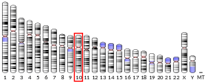
| 위치(UCSC) | Chr 10: 17.23 – 17.24Mb | n/a | |||||||||||||||||||||||
| PubMed 검색 | [2] | [3] | |||||||||||||||||||||||
| 위키다타 | |||||||||||||||||||||||||
| |||||||||||||||||||||||||
비멘틴은 인간에게 VIM 유전자에 의해 암호화된 구조 단백질이다. 이것의 이름은 유연한 막대들의 배열을 가리키는 라틴어 vimentum에서 유래되었다.[4]

비멘틴은 중간 필라멘트(IF) 단백질의 일종으로 중피세포에서 발현된다. 만약 박테리아뿐만 아니라 모든 동물 세포에서[5] 단백질이 발견된다면.[6] 중간 필라멘트는 튜불린 기반의 미세관 및 액틴 기반의 미세섬유와 함께 사이토스켈레톤으로 구성된다. 모든 IF 단백질은 매우 발달적으로 조절되는 방식으로 표현된다; 비멘틴은 중피세포의 주요한 세포골격계 성분이다. 이 때문에 비멘틴은 정상 발육과 전이 과정 모두에서 상피 대 메스꺼움 전이(EMT)를 거치는 중간에서 파생된 세포나 세포의 표지로 자주 사용된다.
구조
다른 모든 중간 필라멘트와 마찬가지로 비멘틴 단량체는 비헬리컬 아미노(머리)와 카복실(꼬리) 영역으로 양 끝을 덮은 중앙 α-헬리컬 영역을 가지고 있다.[7] 두 개의 모노머는 비멘틴 어셈블리의 기본 하위 단위인 코일 코일 다이머 형성을 용이하게 하는 방식으로 공동번역적으로 표현될 가능성이 높다.[8]
α-헬리컬 시퀀스에는 나선의 표면에 "수소 봉인"을 형성하는 데 기여하는 소수성 아미노산 패턴이 포함되어 있다.[7] 또, 코일 코일 디미너를 안정시키는 데 중요한 역할을 하는 것으로 보이는 산성과 기초 아미노산의 주기적인 분포가 있다.[7] 충전된 잔류물의 간격은 α-헬릭스 구조의 안정화가 가능한 이온 염교량에 최적이다. 이러한 유형의 안정화는 체인 간 상호작용이 아니라 차내 상호작용에 직관적이지만, 과학자들은 아마도 산성 및 기본 잔류물로 형성된 차내 염교에서 체인 간 이온 결합으로의 전환이 필라멘트의 조립에 기여한다고 제안했다.[7]
함수
비멘틴은 사이토솔에서 오르가넬의 위치를 지지하고 고정시키는 데 중요한 역할을 한다. 비멘틴은 좌우로 또는 말기로 핵, 소포체 망막, 미토콘드리아에 부착되어 있다.[9]
비멘틴의 동적 특성은 세포에 유연성을 제공할 때 중요하다. 과학자들은 비멘틴이 체내 기계적 스트레스를 받을 때 미세관이나 액틴 필라멘트 네트워크에서 없는 복원력을 세포에 제공한다는 것을 발견했다. 따라서 일반적으로 비멘틴은 세포의 건전성을 유지하는 역할을 하는 세포골격계 성분으로 인정된다. (비멘틴이 없는 세포는 마이크로파괴로 교란되었을 때 극히 섬세하다는 것이 밝혀졌다.)[10] 비멘틴이 부족한 유전자변형 생쥐는 정상으로 나타났고 기능적 차이를 보이지 않았다.[11] 마이크로튜브 네트워크가 중간 네트워크의 부재를 보상했을 가능성이 있다. 이 결과는 마이크로튜브와 비멘틴 사이의 친밀한 상호작용을 지원한다. 더욱이 미세관 탈고화제가 존재했을 때 비멘틴 재구성이 일어나 다시 한 번 두 시스템 간의 관계를 암시했다.[10] 반면에, 비멘틴 유전자가 부족한 상처 입은 쥐들은 야생 쥐들보다 더 느리게 치유된다.[12]
본질적으로 비멘틴은 세포 형태 유지, 세포질의 무결성, 세포골격계 상호작용을 안정화시키는 역할을 한다. 비멘틴은 JUNQ에서 독성 단백질을 제거하고, 아이팟은 포유류 세포 라인의 비대칭 분할에서 독성 단백질을 제거하는 것으로 밝혀졌다.[13]
또한, 비멘틴은 저밀도 지단백질인 LDL의 수송을 통제하는 것으로 밝혀졌으며 - 콜레스테롤을 리소솜에서 에스테르화 현장으로 유도했다.[14] 세포 내 LDL 유래 콜레스테롤의 운반이 차단되면서, 세포는 비멘틴을 함유한 일반 세포보다 훨씬 낮은 비율의 지단백질을 저장하는 것으로 밝혀졌다. 이러한 의존성은 세포 중간 필라멘트 네트워크에 의존하는 어떤 세포에서든 생화학적 기능의 첫 번째 과정인 것 같다. 이러한 형태의 의존은 부신 세포에 영향을 미치며, 부신 세포는 LDL에서 파생된 굴절 에스테르에 의존한다.[14]
비멘틴은 응집된 단백질의 핵심을 둘러싸고 있는 새장을 형성하는 농업생성에 역할을 한다.[15]
임상적 유의성
그것은 중수체를 식별하기 위해 육종 종양 표지로 사용되어 왔다.[16][17] 바이오마커로서의 그것의 특수성은 제라드 가드너에 의해 논란이 되어왔다.[18]
비멘틴 유전자의 메틸화가 대장암의 바이오마커로 정착되어 대장암 대변검사 개발에 활용되고 있다. 바렛의 식도, 식도선두암, 장형위암과 같은 특정 상부위장관 병리학에서도 통계적으로 유의미한 수준의 비멘틴 유전자 메틸화가 관찰되었다.[19] 촉진자 지역의 높은 수준의 DNA 메틸화는 또한 호르몬 양성 유방암에서의 현저한 생존 감소와 관련이 있다.[20] 비멘틴의 하향 조절은 프로테오믹 접근법을 이용한 유두 갑상선암의 낭포성 변종에서 확인되었다.[21] 류마티스 관절염 진단에 사용되는 항시민성 단백질 항체를 참조하십시오.
비멘틴은 네이더 라히미에 의해 사스-CoV-2의 부착 인자로 밝혀졌다.[22]
상호작용
Vimentin은 다음과 상호 작용하는 것으로 나타났다.
비멘틴 mRNA의 3' UTR는 46kDa 단백질을 결합하는 것으로 밝혀졌다.[34]
참조
- ^ a b c GRCh38: 앙상블 릴리스 89: ENSG000026025 - 앙상블, 2017년 5월
- ^ "Human PubMed Reference:". National Center for Biotechnology Information, U.S. National Library of Medicine.
- ^ "Mouse PubMed Reference:". National Center for Biotechnology Information, U.S. National Library of Medicine.
- ^ Franke WW, Schmid E, Osborn M, Weber K (October 1978). "Different intermediate-sized filaments distinguished by immunofluorescence microscopy". Proceedings of the National Academy of Sciences of the United States of America. 75 (10): 5034–8. Bibcode:1978PNAS...75.5034F. doi:10.1073/pnas.75.10.5034. PMC 336257. PMID 368806.
- ^ Eriksson JE, Dechat T, Grin B, Helfand B, Mendez M, Pallari HM, Goldman RD (July 2009). "Introducing intermediate filaments: from discovery to disease". The Journal of Clinical Investigation. 119 (7): 1763–71. doi:10.1172/JCI38339. PMC 2701876. PMID 19587451.
- ^ Cabeen MT, Jacobs-Wagner C (2010). "The bacterial cytoskeleton". Annual Review of Genetics. 44: 365–92. doi:10.1146/annurev-genet-102108-134845. PMID 21047262.
- ^ a b c d Fuchs E, Weber K (1994). "Intermediate filaments: structure, dynamics, function, and disease". Annual Review of Biochemistry. 63: 345–82. doi:10.1146/annurev.bi.63.070194.002021. PMID 7979242.
- ^ Chang L, Shav-Tal Y, Trcek T, Singer RH, Goldman RD (February 2006). "Assembling an intermediate filament network by dynamic cotranslation". The Journal of Cell Biology. 172 (5): 747–58. doi:10.1083/jcb.200511033. PMC 2063706. PMID 16505169.
- ^ Katsumoto T, Mitsushima A, Kurimura T (1990). "The role of the vimentin intermediate filaments in rat 3Y1 cells elucidated by immunoelectron microscopy and computer-graphic reconstruction". Biology of the Cell. 68 (2): 139–46. doi:10.1016/0248-4900(90)90299-I. PMID 2192768. S2CID 29019928.
- ^ a b Goldman RD, Khuon S, Chou YH, Opal P, Steinert PM (August 1996). "The function of intermediate filaments in cell shape and cytoskeletal integrity". The Journal of Cell Biology. 134 (4): 971–83. doi:10.1083/jcb.134.4.971. PMC 2120965. PMID 8769421.
- ^ Colucci-Guyon E, Portier MM, Dunia I, Paulin D, Pournin S, Babinet C (November 1994). "Mice lacking vimentin develop and reproduce without an obvious phenotype". Cell. 79 (4): 679–94. doi:10.1016/0092-8674(94)90553-3. PMID 7954832. S2CID 28146121.
- ^ Eckes B, Colucci-Guyon E, Smola H, Nodder S, Babinet C, Krieg T, Martin P (July 2000). "Impaired wound healing in embryonic and adult mice lacking vimentin". Journal of Cell Science. 113 (13): 2455–62. doi:10.1242/jcs.113.13.2455. PMID 10852824.
- ^ Ogrodnik M, Salmonowicz H, Brown R, Turkowska J, Średniawa W, Pattabiraman S, Amen T, Abraham AC, Eichler N, Lyakhovetsky R, Kaganovich D (June 2014). "Dynamic JUNQ inclusion bodies are asymmetrically inherited in mammalian cell lines through the asymmetric partitioning of vimentin". Proceedings of the National Academy of Sciences of the United States of America. 111 (22): 8049–54. Bibcode:2014PNAS..111.8049O. doi:10.1073/pnas.1324035111. PMC 4050583. PMID 24843142.
- ^ a b Sarria AJ, Panini SR, Evans RM (September 1992). "A functional role for vimentin intermediate filaments in the metabolism of lipoprotein-derived cholesterol in human SW-13 cells". The Journal of Biological Chemistry. 267 (27): 19455–63. doi:10.1016/S0021-9258(18)41797-8. PMID 1527066.
- ^ Johnston JA, Ward CL, Kopito RR (December 1998). "Aggresomes: a cellular response to misfolded proteins". The Journal of Cell Biology. 143 (7): 1883–98. doi:10.1083/jcb.143.7.1883. PMC 2175217. PMID 9864362.
- ^ Leader M, Collins M, Patel J, Henry K (January 1987). "Vimentin: an evaluation of its role as a tumour marker". Histopathology. 11 (1): 63–72. doi:10.1111/j.1365-2559.1987.tb02609.x. PMID 2435649. S2CID 34804720.
- ^ "Immunohistochemistry from the Washington Animal Disease Diagnostic laboratory (WADDL)of the College of Veterinary Medicine, Washington State University". Archived from the original on 2008-12-01. Retrieved 2009-03-14.
- ^ Gardner, Jerad (23 September 2010). "How to Interpret Vimentin Immunostain". YouTube. Archived from the original on 2021-12-12.
- ^ Moinova H, Leidner RS, Ravi L, Lutterbaugh J, Barnholtz-Sloan JS, Chen Y, Chak A, Markowitz SD, Willis JE (April 2012). "Aberrant vimentin methylation is characteristic of upper gastrointestinal pathologies". Cancer Epidemiology, Biomarkers & Prevention. 21 (4): 594–600. doi:10.1158/1055-9965.EPI-11-1060. PMC 3454489. PMID 22315367.
- ^ Ulirsch J, Fan C, Knafl G, Wu MJ, Coleman B, Perou CM, Swift-Scanlan T (January 2013). "Vimentin DNA methylation predicts survival in breast cancer". Breast Cancer Research and Treatment. 137 (2): 383–96. doi:10.1007/s10549-012-2353-5. PMC 3838916. PMID 23239149.
- ^ Dinets A, Pernemalm M, Kjellin H, Sviatoha V, Sofiadis A, Juhlin CC, Zedenius J, Larsson C, Lehtiö J, Höög A (2015). "Differential protein expression profiles of cyst fluid from papillary thyroid carcinoma and benign thyroid lesions". PLOS ONE. 10 (5): e0126472. Bibcode:2015PLoSO..1026472D. doi:10.1371/journal.pone.0126472. PMC 4433121. PMID 25978681.
- ^ 암레이, R, 샤아, C, 올레즈니크, J, 화이트, M.R., 나폴레옹, M.A., 로트폴라자데, S. 하우저, B.M., 슈미트, A.G., 치탈리아, V., 뮐버거, E. 외(2022년). 세포외 비멘틴은 SARS-CoV-2의 인체 내피세포 진입을 용이하게 하는 부착인자다. Natl Acad Sci U S 119를 신청하십시오.
- ^ Meng JJ, Bornslaeger EA, Green KJ, Steinert PM, Ip W (August 1997). "Two-hybrid analysis reveals fundamental differences in direct interactions between desmoplakin and cell type-specific intermediate filaments". The Journal of Biological Chemistry. 272 (34): 21495–503. doi:10.1074/jbc.272.34.21495. PMID 9261168.
- ^ Lopez-Egido J, Cunningham J, Berg M, Oberg K, Bongcam-Rudloff E, Gobl A (August 2002). "Menin's interaction with glial fibrillary acidic protein and vimentin suggests a role for the intermediate filament network in regulating menin activity". Experimental Cell Research. 278 (2): 175–83. doi:10.1006/excr.2002.5575. PMID 12169273.
- ^ Rual JF, Venkatesan K, Hao T, Hirozane-Kishikawa T, Dricot A, Li N, et al. (October 2005). "Towards a proteome-scale map of the human protein-protein interaction network". Nature. 437 (7062): 1173–8. Bibcode:2005Natur.437.1173R. doi:10.1038/nature04209. PMID 16189514. S2CID 4427026.
- ^ Stelzl U, Worm U, Lalowski M, Haenig C, Brembeck FH, Goehler H, et al. (September 2005). "A human protein-protein interaction network: a resource for annotating the proteome". Cell. 122 (6): 957–68. doi:10.1016/j.cell.2005.08.029. hdl:11858/00-001M-0000-0010-8592-0. PMID 16169070. S2CID 8235923.
- ^ Matsuzawa K, Kosako H, Inagaki N, Shibata H, Mukai H, Ono Y, et al. (May 1997). "Domain-specific phosphorylation of vimentin and glial fibrillary acidic protein by PKN". Biochemical and Biophysical Research Communications. 234 (3): 621–5. doi:10.1006/bbrc.1997.6669. PMID 9175763.
- ^ Ratnayake WS, Apostolatos AH, Ostrov DA, Acevedo-Duncan M (November 2017). "Two novel atypical PKC inhibitors; ACPD and DNDA effectively mitigate cell proliferation and epithelial to mesenchymal transition of metastatic melanoma while inducing apoptosis". International Journal of Oncology. 51 (5): 1370–1382. doi:10.3892/ijo.2017.4131. PMC 5642393. PMID 29048609.
- ^ Ratnayake WS, Apostolatos CA, Apostolatos AH, Schutte RJ, Huynh MA, Ostrov DA, Acevedo-Duncan M (2018). "Oncogenic PKC-ι activates Vimentin during epithelial-mesenchymal transition in melanoma; a study based on PKC-ι and PKC-ζ specific inhibitors". Cell Adhesion & Migration. 12 (5): 447–463. doi:10.1080/19336918.2018.1471323. PMC 6363030. PMID 29781749.
- ^ Herrmann H, Wiche G (January 1987). "Plectin and IFAP-300K are homologous proteins binding to microtubule-associated proteins 1 and 2 and to the 240-kilodalton subunit of spectrin". The Journal of Biological Chemistry. 262 (3): 1320–5. doi:10.1016/S0021-9258(19)75789-5. PMID 3027087.
- ^ a b Brown MJ, Hallam JA, Liu Y, Yamada KM, Shaw S (July 2001). "Cutting edge: integration of human T lymphocyte cytoskeleton by the cytolinker plectin". Journal of Immunology. 167 (2): 641–5. doi:10.4049/jimmunol.167.2.641. PMID 11441066.
- ^ Russell RL, Cao D, Zhang D, Handschumacher RE, Pizzorno G (April 2001). "Uridine phosphorylase association with vimentin. Intracellular distribution and localization". The Journal of Biological Chemistry. 276 (16): 13302–7. doi:10.1074/jbc.M008512200. PMID 11278417.
- ^ Tzivion G, Luo ZJ, Avruch J (September 2000). "Calyculin A-induced vimentin phosphorylation sequesters 14-3-3 and displaces other 14-3-3 partners in vivo". The Journal of Biological Chemistry. 275 (38): 29772–8. doi:10.1074/jbc.M001207200. PMID 10887173.
- ^ Zehner ZE, Shepherd RK, Gabryszuk J, Fu TF, Al-Ali M, Holmes WM (August 1997). "RNA-protein interactions within the 3 ' untranslated region of vimentin mRNA". Nucleic Acids Research. 25 (16): 3362–70. doi:10.1093/nar/25.16.3362. PMC 146884. PMID 9241253.
추가 읽기
- Snásel J, Pichová I (1997). "The cleavage of host cell proteins by HIV-1 protease". Folia Biologica. 42 (5): 227–30. doi:10.1007/BF02818986. PMID 8997639. S2CID 7617882.
- Lake JA, Carr J, Feng F, Mundy L, Burrell C, Li P (February 2003). "The role of Vif during HIV-1 infection: interaction with novel host cellular factors". Journal of Clinical Virology. 26 (2): 143–52. doi:10.1016/S1386-6532(02)00113-0. PMID 12600646.


