망상형성
Reticular formation| 망상형성 | |
|---|---|
 | |
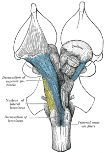
 올리브의 중간쯤에 있는 수근의 횡단부(왼쪽의 망상망막과 망상망막 알바) | |
| 세부 사항 | |
| 위치 | 뇌간 |
| 식별자 | |
| 라틴어 | 망막형성 |
| 메쉬 | D012154 |
| 신경명 | 1223 |
| NeuroLex ID | nlx_143558 |
| TA98 | A14.1.00.021 A14.1.05.403 A14.1.06.327 |
| TA2 | 5367 |
| FMA | 77719 |
| 신경해부술의 해부학적 용어 | |
망상형성은 뇌간 전체에 위치한 서로 연결된 핵의 집합이다.이것은 뇌의 다른 부분에 위치한 뉴런을 포함하고 있기 때문에 해부학적으로 잘 정의되어 있지 않다.망상 형성의 뉴런은 뇌간 중심부에서 중뇌의 상부에서 수질의 [2]하부에 이르는 복잡한 네트워크 세트를 형성합니다.망상형성은 상행망막활성화계(ARAS)의 피질에 대한 상승경로와 망상척수관을 [3][4][5][6]통한 척수에 대한 하강경로를 포함한다.
망상형성의 뉴런, 특히 상승망상활성계의 뉴런은 행동적인 각성과 의식을 유지하는 데 중요한 역할을 한다.망상 형성의 전반적인 기능은 체세포 운동 제어, 심혈관 제어, 통증 조절, 수면과 의식, 그리고 [7]습관화를 포함하는 조절과 [A]전운동이다.조절 기능은 주로 망상 형성의 로스트랄 부분에서 발견되며, 전운동 기능은 더 많은 꼬리 부분의 뉴런에서 국소화된다.
망상형성은 세 개의 열로 나뉜다: 라페핵(중간), 기간토세포망상핵(중간대), 파보세포망상핵(중간대)라페핵은 기분 조절에 중요한 역할을 하는 신경전달물질 세로토닌의 합성 장소이다.기간토 세포핵은 운동 협응에 관여한다.파세포핵은 [8]호흡을 조절한다.
망상 형성은 고등 유기체의 기본 기능 중 일부를 통제하는데 필수적이며 뇌의 [citation needed]계통학적으로 가장 오래된 부분 중 하나입니다.
구조.

인간의 망상 형성은 거의 100개의 뇌핵으로 구성되어 있으며 다른 영역들 [3]중에서 전뇌, 뇌간, 소뇌에 많은 돌기를 포함합니다.망상핵,[B] 망상 시상피질투영섬유, 확산성 시상피질투영, 상승콜린성투영, 하강비콜린성투영 및 하강망상척수투영 [4]등이다.망상형성은 또한 두 개의 주요 신경 서브시스템, 즉 뚜렷한 인지적 및 [3][4]생리학적 과정을 매개하는 상행망상 활성화 시스템과 하행망상척수관을 포함합니다.그것은 궁수와 관상 양쪽에서 기능적으로 갈라져 있다.
전통적으로 망상핵은 세 개의 열로 나뉩니다.
- 중앙 열 – 라페 핵
- 중앙 열 – 기가토 세포 핵(세포의 크기가 크기 때문에)
- 측면 기둥 – 파세포 핵(세포의 크기가 작기 때문에)
원래의 기능적 차별화는 꼬리뼈와 로스트랄의 분할이었다.이는 늑골망막형성의 병변이 고양이 뇌에서 과민증을 유발한다는 관찰에 기초했다.이와는 대조적으로, 망상 형성의 더 많은 꼬리 부분의 병변은 고양이에서 불면증을 일으킨다.이 연구는 꼬리 부분이 망상 형성의 로스트랄 부분을 억제한다는 생각으로 이어졌다.
시상 분열은 더 많은 형태학적 차이를 드러낸다.라페 핵은 망상 형성 중간에 융기를 형성하고, 그 주변에는 내측 망상 형성이라고 불리는 분할이 있습니다.내측 RF는 크고 긴 상승 및 하강 섬유를 가지고 있으며, 외측 망막 형성으로 둘러싸여 있습니다.측면 RF는 두개골 신경의 운동핵에 가깝고 대부분 그 기능을 중개합니다.
내측 및 외측 망상 형성
내측망막형성과 외측망막형성은 수질을 통해 중뇌로 돌출부를 보내는 경계가 명확하지 않은 두 개의 핵 기둥이다.핵은 기능, 세포 유형, 그리고 효모 또는 구심성 신경의 돌기에 의해 구별될 수 있다.로스트랄 중뇌에서 후두부로 이동하면 로스트랄 종아리 부위와 중뇌 부위에서 내측 RF가 덜 두드러지고 측면 RF가 [citation needed]더 두드러진다.
내측망막형성의 측면에는 외측사촌이 존재하며, 외측사촌은 특히 로스트랄수골과 꼬리뼈에서 두드러진다.이 부위에서 매우 중요한 [clarification needed]미주신경을 포함한 두개신경이 튀어나온다.측면 RF는 뇌신경 주변의 신경절과 인터뉴론의 영역으로 알려져 있으며, 이는 그들의 특징적인 반사와 기능을 중재하는 역할을 한다.
기능.
망상형성은 다음을 포함한 다양한 기능을 가진 100개 이상의 작은 신경망으로 구성됩니다.
- 체세포 운동 제어 – 일부 운동 뉴런은 축삭을 망상 형성 핵으로 보내 척수의 망상척수관을 발생시킵니다.이 기관들은 특히 몸을 움직이는 동안 톤, 균형, 자세를 유지하는 기능을 합니다.망상 형성 또한 소뇌가 시각, 청각, 전정 자극을 운동 협응에 통합할 수 있도록 눈과 귀 신호를 소뇌에 전달합니다.다른 운동핵에는 시선이 물체를 추적하고 고정할 수 있는 시선중추와 호흡과 삼키는 리듬 신호를 생성하는 중앙 패턴 발생기가 있습니다.
- 심혈관 제어 – 망상 형성에는 수막의 심장 및 혈관 운동 중심이 포함됩니다.
- 통증 조절 – 망상 형성은 하체의 통증 신호가 대뇌 피질에 도달하는 수단 중 하나입니다.그것은 또한 하강 진통 경로의 기원이다.이러한 경로의 신경 섬유는 뇌로 전달되는 통증 신호를 차단하기 위해 척수에서 작용합니다.
- 수면과 의식 – 망상 형성은 어떤 감각 신호가 대뇌에 도달하고 우리의 의식적인 주의를 끌 수 있도록 하는 시상과 대뇌 피질에 투영되어 있습니다.그것은 경각심과 수면과 같은 의식 상태에서 중심적인 역할을 한다.망상 형성에 손상을 입으면 돌이킬 수 없는 혼수 상태가 될 수 있습니다.
- 습관 – 이것은 뇌가 다른 사람들에게 민감하게 유지하면서 반복적이고 무의미한 자극을 무시하는 것을 배우는 과정이다.대도시의 교통소음 속에서도 잠을 잘 수 있지만 알람 소리나 아기 울음소리에 잠을 깨는 사람이 그 좋은 예다.대뇌피질의 활동을 조절하는 망상형성핵은 상승망상활성계의 일부이다.[9][7]
메이저 서브시스템
상행망막활성화계
상행망막활성화계(ARAS)는 시상외조절계 또는 간단히 망막활성화계(RAS)로도 알려져 있으며, 각성과 수면-각성 전환을 조절하는 척추동물의 뇌에서 연결된 핵 세트이다.ARAS는 망상 형성의 일부이며 대부분 시상 내의 다양한 핵과 도파민 작동성, 노르아드레날린 작동성, 세로토닌 작동성, 히스타민 작동성, 콜린 작동성 및 글루탐산성 [3][10][11][12]뇌핵으로 구성되어 있습니다.
ARAS의 구조
ARAS는 시상과 [3][11][12]시상하부를 통해 돌출된 뚜렷한 경로를 통해 대뇌피질에 후미 중뇌와 전두엽의 등부를 연결하는 여러 개의 신경 회로로 구성되어 있습니다.ARAS는 서로 다른 핵들의 집합입니다 – 뇌간 상층부, 뇌간, 뇌수, 시상하부의 각 측면에 각각 20개 이상 있습니다.이 뉴런들이 방출하는 신경전달물질은 도파민, 노르에피네프린, 세로토닌, 히스타민, 아세틸콜린,[3][10][11][12] 글루탐산염을 포함한다.이들은 시상 릴레이를 [11][12][13]통한 직접 축방향 투영과 간접 투영을 통해 피질적 영향을 미친다.
시상 경로는 주로 폰틴 테그넘의 콜린 작동성 뉴런으로 구성되고 시상하부 경로는 주로 도파민, 노르에피네프린, 세로토닌, 히스타민 등 모노아민 신경전달물질을 [3][10]방출하는 뉴런으로 구성됩니다.ARAS의 글루탐산 방출 뉴런은 모노아민성 및 콜린성 [14]핵과 관련하여 훨씬 최근에 확인되었다. ARAS의 글루탐산 성분은 시상하부의 하나의 핵과 다양한 뇌간 [11][14][15]핵을 포함한다.시상하부의 외측 신경 세포는 상승망막 활성화 시스템의 모든 구성 요소를 형성하고 전체 [12][16][17]시스템 내에서 좌표 활동을 조절합니다.
| 핵형 | 각성을 매개하는 해당 핵 | 원천 |
|---|---|---|
| 도파민성 핵 | [3][10][11][12] | |
| 노르아드레날린 핵 |
| [3][10][12] |
| 세로토닌성 핵 | [3][10][12] | |
| 히스타민 작동성 핵 | [3][10][18] | |
| 콜린 작동성 핵 |
| [3][11][12][14] |
| 글루탐산성 핵 |
| [11][12][14][15][18][19] |
| 시상핵 | [3][11][20] |
ARAS는 동물의 생존에 중요하며 동물의 [C][22]최면이라고 불리는 토셀레플렉스의 억제 기간과 같은 불리한 기간 동안 보호되는 뇌의 진화적으로 오래된 영역으로 구성됩니다.신경조절돌기를 피질에 보내는 상행망막활성화계는 주로 전전두피질에 [23]연결된다.대뇌피질의 [23]운동부위와의 연결성이 낮은 것 같습니다.
ARAS의 기능
의식
상승망막 활성화 시스템은 의식 [13]상태를 가능하게 하는 중요한 요소이다.상승 시스템은 피질적 및 행동적 [6]각성으로 특징지어지는 각성에 기여하는 것으로 보인다.
sleep-wake 전환 조절
ARAS의 주요 기능은 시상 및 피질 기능을 수정하고 강화하여 뇌파(EEG) 비동기화가 [D][25][26]뒤따르도록 하는 것입니다.각성 및 수면 기간 동안 뇌의 전기 활동에는 뚜렷한 차이가 있습니다: 저전압의 빠른 뇌파(EEG 비동기화)는 각성 및 렘 수면과 관련이 있으며, 고전압의 느린 파동은 비렘 수면 중에 발견됩니다.일반적으로 시상 릴레이 뉴런이 버스트 모드일 때는 EEG가 동기화되고 강장 모드일 때는 [26]비동기화됩니다.ARAS의 자극은 느린 피질파(0.3~1Hz), 델타파(1~4Hz), 스핀들파 진동(11~14Hz)을 억제하고 감마밴드(20~40Hz) 발진을 [16]촉진함으로써 EEG 비동기화를 생성합니다.
깊은 수면 상태에서 깨어 있는 상태로의 생리적인 변화는 ARAS에 [27]의해 가역적이고 중재된다.시상하부의 복측전안핵(VLPO)은 각성상태를 담당하는 신경회로를 억제하고 VLPO 활성화는 수면개시에 [28]기여한다.수면 중에 ARAS의 뉴런은 훨씬 낮은 발화율을 가질 것이고, 반대로, 그들은 깨어 있는 [29]동안 더 높은 활동 수준을 가질 것입니다.뇌가 잠을 자려면 ARAS의 [27]억제에 의해 피질에 도달하는 상승 구심성 활동이 감소해야 한다.
주의
ARAS는 또한 편안한 각성 상태에서 높은 [20]주의 기간으로의 전환을 중재하는 데 도움이 됩니다.주의력과 주의가 필요한 과제 동안 중뇌망막형성(MRF)과 시상내핵에서 국소 혈류 증가(아마도 신경 활동의 증가를 나타냄)가 있다.
ARAS의 임상적 의의
뇌간 ARAS 핵의 집단 병변은 의식 수준에 심각한 변화를 일으킬 수 있다(예: 혼수).[30]중뇌의 망상 형성에 대한 양쪽 손상은 혼수 또는 [31]사망으로 이어질 수 있다.
ARAS의 직접적인 전기적 자극은 고양이에게 통증 반응을 일으키고 인간의 [citation needed]통증에 대한 구두 보고를 유도한다.고양이의 상승성 망막 활성화는 지속적인 통증으로 인해 발생할 수 있는 산드라이아시스를 [32]발생시킬 수 있다.이러한 결과는 ARAS 회로와 생리적인 통증 [32]경로 사이의 관계를 시사한다.
병리
나이가 들수록 [33]ARAS의 반응성이 전반적으로 저하되기 때문에 ARAS의 병리 중 일부는 연령에 기인할 수 있다.전기[E] 커플링의 변화는 ARAS 활동의 일부 변화를 설명하기 위해 제안되었다. 커플링이 다운 조절된 경우 고주파 동기(감마 밴드)에 상응하는 감소가 있을 것이다.반대로, 상향 조절 전기 커플링은 각성 및 REM 수면 [35]구동의 증가를 초래할 수 있는 빠른 리듬의 동기화를 증가시킬 것이다.특히 ARAS의 중단은 다음 장애와 관련이 있습니다.
- 기면증:Pedunculopontine (PPT/PN) / Laterodorsal teg멘탈 (LDT)핵을 따른 병변은 기면증과 [36]관련이 있다.PPN 생산량의 유의한 하향 조절과 오렉신 펩타이드의 손실이 있어 이 [16]질환의 특징인 과도한 주간 졸음을 촉진한다.
- 진행성 핵상마비(PSP): 아산화질소 시그널링의 기능 부전이 PSP의 [37]발달에 관여하고 있다.
- 파킨슨병: 렘수면장애는 파킨슨병에서 흔하다.주로 도파민성 질환이지만 콜린성 핵도 고갈되어 있습니다.ARAS의 변성은 질병 [36]과정 초기에 시작된다.
발달상의 영향
상승망막 활성화 시스템의 발달에 악영향을 미칠 수 있는 몇 가지 잠재적 요인이 있습니다.
- 조산:[38] 출생 체중이나 임신 주수에 관계없이, 조산은 발달 내내 주의력, 주의력, 그리고 피질 메커니즘에 지속적인 유해한 영향을 유발합니다.
- 임신 [39]중 흡연: 태아에서 담배 연기에 노출되는 것은 인간에게 지속적인 각성, 주의력 및 인지적 결함을 발생시키는 것으로 알려져 있습니다.이러한 노출은 pedunculopontine nucleus(PN) 세포에서 α4β2 니코틴 수용체의 상향 조절을 유도하여 강장 활성, 휴지막 전위 및 과분극 활성화 카티온 전류를 발생시킬 수 있다.PPN 뉴런의 내재막 특성에 대한 이러한 주요 장애는 각성 및 감각 동기, 결손의 증가 수준을 초래한다(반복적인 청각 자극에 대한 습관 감소로 입증됨).이러한 생리학적 변화가 나중에 주의력 저하를 심화시킬 수 있다는 가설이 있다.
내림막척수관
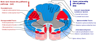
하강 또는 전방 망상척수관이라고도 알려진 망상척수관은 두 관점의 망상형성으로부터[40] 내려오는 추체외 운동관이며, 간선 및 근위 사지 굴곡 및 신장을 공급하는 운동신경세포에 작용합니다.망상척수관은 다른 기능도 있지만 주로 이동과 자세 제어에 [41]관여한다.하행 망상척수관은 근골격계 활동을 위해 척수로 가는 4가지 주요 피질 경로 중 하나이다.망상척수관은 다른 세 가지 경로와 함께 작동하여 섬세한 [40]조작을 포함한 움직임을 조정 제어한다.네 가지 경로는 두 가지 주요 시스템 경로, 즉 중간 시스템과 측면 시스템으로 분류할 수 있다.내측 시스템은 망상척수 경로와 전정척수 경로를 포함하며, 이 시스템은 자세의 제어를 제공한다.피질척수 및 루브로척수관 경로는 움직임을 [40]미세하게 제어하는 측면 시스템에 속합니다.
망상척수관 구성 요소
이 하강관은 내측(또는 폰틴)과 외측(또는 수) 망상척수관(MRST 및 LRST)의 두 부분으로 나뉩니다.
- MRST는 자극적인 반중력, 신장근육을 담당한다.이 관의 섬유는 꼬리 폰틴 망상핵과 구강 폰틴 망상핵에서 발생하며 척수의 라미나 VII와 라미나 VII로 돌출된다.
- LRST는 자극성 축신근 운동 억제를 담당합니다.또한 자동 호흡도 담당합니다.이 관의 섬유는 주로 기간세포핵에서 수질의 망상형성으로부터 발생하며, 측주의 앞부분에서 척수의 길이를 내려갑니다.이 관은 대부분 척수의 라미나 IX로 끝나는 섬유로 끝납니다.
반대방향으로 정보를 전달하는 상승감각로를 스피노레틱로라고 한다.
망상척수관의 기능
- 모터 시스템의 정보를 통합하여 이동 및 자세의 자동 이동을 조정합니다.
- 자발적인 움직임을 촉진 및 억제하고 근육 긴장도에 영향을 줍니다.
- 자율 기능을 중개합니다.
- 통증 자극을 조절합니다.
- 시상 [42]외측 관절핵으로의 혈류에 영향을 줍니다.
망상척수관의 임상적 의미
망상척수관은 시상하부가 교감성 흉곽유출과 [citation needed]부교감성 천골유출을 제어할 수 있는 경로를 제공한다.
뇌간과 소뇌에서 척수로 신호를 전달하는 두 가지 주요 하강 시스템은 균형과 방향을 위한 자동 자세 반응을 촉발할 수 있습니다: 전정핵으로부터의 전정척수관로 및 종아리 및 수질로부터의 망막척수관로입니다.이 기관들의 병변은 심각한 운동실조와 자세의 [43]불안정성을 초래한다.
적핵(중뇌)과 전정핵(전정핵)을 분리하는 뇌간의 물리적 또는 혈관 손상은 근육 긴장 증가와 과잉 활동적 스트레치 반사의 신경학적 징후를 보이는 뇌경직을 일으킬 수 있다.놀랍거나 고통스러운 자극에 반응하여 팔과 다리는 내부를 향해 뻗고 회전합니다.원인은 루브척수관의 억제 없이 신장운동신경계를 자극하는 외측전정척수 및 망상척수관의 [44]강장활동이다.
뇌간 손상이 붉은 핵을 초과하면 피질 경직이 발생할 수 있습니다.놀랍거나 고통스러운 자극에 반응하여 팔은 구부리고 다리는 뻗는다.원인은 외측 전정척수관 및 망상척수관에서 신전척수관의 들뜸을 상쇄하는 루브로척수관을 통해 붉은 핵이다.루브척수관은 경추까지만 확장되기 때문에 [44]다리보다는 굴곡근육을 자극하고 신장을 억제함으로써 대부분 팔에 작용한다.
전정핵 아래 수질에 손상이 생기면 박리성 마비, 저혈압, 호흡동력 상실, 사지마비 등을 일으킬 수 있다.외측 전정척수 [44]및 망상척수기관에서 발생하는 강장활동이 더 이상 없기 때문에 운동신경계의 완전한 활동 상실로 인해 척추 쇼크의 초기 단계와 유사한 반사가 없다.
역사
"망막 형성"이라는 용어는 19세기 후반 오토 디테르에 의해 라몬 이 카잘의 뉴런 원칙과 일치하면서 만들어졌습니다.앨런 홉슨은 그의 저서 망상형성(The Reticular Formation Reversited)에서 그 이름은 신경과학의 집합장 이론이 몰락한 시대의 어원적 흔적이라고 언급하고 있다."망상"이란 "망상 구조"를 의미하며, 망상 형성은 언뜻 보기에 이와 유사하다.그것은 연구하기에는 너무 복잡하거나 전혀 조직이 없는 뇌의 분화되지 않은 부분으로 묘사되어 왔다.Eric Kandel은 망상 형성이 척수의 중간 회백질과 유사한 방식으로 구성되었다고 설명합니다.이 혼란스럽고, 느슨하고, 복잡한 형태의 조직은 많은 연구자들이 뇌의 [citation needed]특정 영역을 더 멀리 들여다보는 것을 꺼리게 만들었다.세포는 명확한 신경절 경계가 없지만, 분명한 기능적 조직과 뚜렷한 세포 유형을 가지고 있습니다."망막 형성"이라는 용어는 일반론을 말하는 것 외에는 더 이상 거의 사용되지 않는다.현대의 과학자들은 보통 망상 [citation needed]형성을 구성하는 개별 핵을 말한다.
Moruzi와 Magoun은 1949년에 처음으로 뇌의 수면-깨우기 메커니즘을 조절하는 신경 성분을 조사했다.생리학자들은 뇌의 깊은 곳에 있는 어떤 구조가 정신적인 각성과 [25]경각심을 조절한다고 제안했었다.각성은 대뇌피질에서 구심성(감각성) 자극의 직접적인 수신에만 의존한다고 여겨져 왔다.
뇌의 직접적인 전기 자극이 전기 피질 계전기를 시뮬레이션 할 수 있기 때문에, Magoun은 이 원리를 고양이의 뇌간 두 개의 분리된 영역에서 잠에서 깨는 방법을 시연하기 위해 사용했습니다.그는 첫 번째로 상승하는 체력과 청각 경로를 자극했다; 두 번째로, "하뇌의 망상 형성에서 상승하는 계전기는 중뇌 테그넘, 시상하부를 통해 내부 [45]캡슐로 이어진다."이 계전기는 각성 신호 전달을 위한 알려진 해부학적 경로에 해당하지 않고 상승망막활성화시스템(ARAS)으로 명명되었기 때문에 특히 관심을 끌었다.
다음으로, 새롭게 확인된 릴레이 시스템의 중요성은 중뇌 전면의 안쪽과 측면 부분에 병변을 배치하여 평가하였다.ARAS에 대한 간뇌 간섭이 있는 고양이는 깊은 잠에 들어가 그에 상응하는 뇌파를 보였다.다른 방법으로는, 상승 청각 및 체세포 경로에 유사한 방해를 받은 고양이는 정상적인 수면과 각성을 보였으며 물리적 자극으로 깨어날 수 있었다.이러한 외부 자극이 간섭에 의해 피질로 가는 도중에 차단되기 때문에, 이것은 상승 전달이 새로 발견된 ARAS를 통해 이동해야 한다는 것을 나타냅니다.
마지막으로, Magoun은 뇌간 안쪽 부분의 전위를 기록했고 청각 자극이 망막 활성화 시스템의 일부를 직접 작동시킨다는 것을 발견했습니다.또한 좌골신경의 단일충격 자극은 내측망막형성, 시상하부 및 시상부도 활성화시켰다.ARAS의 들뜸은 소뇌 회로를 통한 추가적인 신호 전파에 의존하지 않았다. 왜냐하면 소뇌와 질식 제거 후에 동일한 결과를 얻었기 때문이다.연구진은 중뇌망형성을 둘러싼 세포기둥이 뇌간의 모든 상승 경로로부터 입력을 받아 이러한 구심성을 피질에 전달하고 따라서 [45][27]각성을 조절하는 것을 제안했다.
「 」를 참조해 주세요.
각주
- ^ 전운동은 피드백 감각 신호를 상부 운동 뉴런과 깊은 소뇌 핵으로부터의 명령과 통합하고, 뇌간과 [2]척수에 하부 내장 운동과 일부 체세포 운동 뉴런의 효율적인 활동을 조직하는 것과 같은 기능을 합니다.
- ^ 수질, 종아리, [4]중뇌의 구조를 포함한 망상핵,
- ^ 동물 최면이란 인간이 아닌 동물에서 운동 반응이 없는 상태를 말한다.이 상태는 쓰다듬기, 두드러진 자극 또는 신체적 구속 때문에 발생할 수 있습니다.그 이름은 인간의 최면술과 [21]무아지경의 유사성 때문에 유래되었다.
- ^ 두피에 있는 EEG의 전극은 기초 뇌 영역에 있는 매우 많은 피라미드형 뉴런의 활동을 측정합니다.각각의 뉴런은 시간이 지남에 따라 변화하는 작은 전기장을 생성한다.수면 상태에서 뉴런은 거의 동시에 활성화되고, 뉴런의 전기장의 합계를 나타내는 EEG파는 위상이 일치하고 진폭이 커지기 때문에 '동기화'된다.깨어 있는 상태에서는 불규칙하거나 위상 외 입력으로 인해 동시에 활성화되지 않으며, 대수 합계를 나타내는 EEG 파형은 더 작은 진폭을 가지므로 "비동기화"[24]됩니다.
- ^ 전기적 결합은 심장 근육의 세포나 전기적 시냅스를 가진 뉴런과 같은 갭 접합을 통해 하나의 세포에서 인접한 세포로 전류가 수동적으로 흐르는 것입니다.전기적으로 결합된 셀은 한 셀에서 발생한 전류가 다른 [34]셀로 빠르게 퍼지기 때문에 동시에 작동한다.
레퍼런스
- ^ Gray, Henry. "FIG. 701: Henry Gray (1825-1861). Anatomy of the Human Body. 1918". Bartleby.com. Archived from the original on 2018-04-21. Retrieved 2019-09-12.
- ^ a b Purves, Dale (2011). Neuroscience (5. ed.). Sunderland, Mass.: Sinauer. pp. 390–395. ISBN 978-0-87893-695-3.
- ^ a b c d e f g h i j k l m Iwańczuk W, Guźniczak P (2015). "Neurophysiological foundations of sleep, arousal, awareness and consciousness phenomena. Part 1". Anaesthesiol Intensive Ther. 47 (2): 162–167. doi:10.5603/AIT.2015.0015. PMID 25940332.
The ascending reticular activating system (ARAS) is responsible for a sustained wakefulness state. It receives information from sensory receptors of various modalities, transmitted through spinoreticular pathways and cranial nerves (trigeminal nerve — polymodal pathways, olfactory nerve, optic nerve and vestibulocochlear nerve — monomodal pathways). These pathways reach the thalamus directly or indirectly via the medial column of reticular formation nuclei (magnocellular nuclei and reticular nuclei of pontine tegmentum). The reticular activating system begins in the dorsal part of the posterior midbrain and anterior pons, continues into the diencephalon, and then divides into two parts reaching the thalamus and hypothalamus, which then project into the cerebral cortex (Fig. 1). The thalamic projection is dominated by cholinergic neurons originating from the pedunculopontine tegmental nucleus of pons and midbrain (PPT) and laterodorsal tegmental nucleus of pons and midbrain (LDT) nuclei [17, 18]. The hypothalamic projection involves noradrenergic neurons of the locus coeruleus (LC) and serotoninergic neurons of the dorsal and median raphe nuclei (DR), which pass through the lateral hypothalamus and reach axons of the histaminergic tubero-mamillary nucleus (TMN), together forming a pathway extending into the forebrain, cortex and hippocampus. Cortical arousal also takes advantage of dopaminergic neurons of the substantia nigra (SN), ventral tegmenti area (VTA) and the periaqueductal grey area (PAG). Fewer cholinergic neurons of the pons and midbrain send projections to the forebrain along the ventral pathway, bypassing the thalamus [19, 20].
- ^ a b c d Augustine JR (2016). "Chapter 9: The Reticular Formation". Human Neuroanatomy (2nd ed.). John Wiley & Sons. pp. 141–153. ISBN 9781119073994. Archived from the original on 4 May 2018. Retrieved 4 September 2017.
- ^ "the definition of reticular activating system". Dictionary.com. Archived from the original on 2017-02-05.
- ^ a b Jones, BE (2008). "Modulation of cortical activation and behavioral arousal by cholinergic and orexinergic systems". Annals of the New York Academy of Sciences. 1129 (1): 26–34. Bibcode:2008NYASA1129...26J. doi:10.1196/annals.1417.026. PMID 18591466. S2CID 16682827.
- ^ a b Saladin, KS (2018). "Chapter 14 - The Brain and Cranial Nerves". Anatomy and Physiology: The Unity of Form and Function (8th ed.). New York: McGraw-Hill. The Reticular Formation, pp. 518-519. ISBN 978-1-259-27772-6.
- ^ "The Brain From Top To Bottom". Thebrain.mcgill.ca. Archived from the original on 2016-04-23. Retrieved 2016-04-28.
- ^ "Anatomy of the Brain - Reticular Formation". Biology.about.com. 2015-07-07. Archived from the original on 2003-04-14. Retrieved 2016-04-28.
- ^ a b c d e f g Malenka RC, Nestler EJ, Hyman SE (2009). "Chapter 12: Sleep and Arousal". In Sydor A, Brown RY (eds.). Molecular Neuropharmacology: A Foundation for Clinical Neuroscience (2nd ed.). New York, USA: McGraw-Hill Medical. p. 295. ISBN 9780071481274.
The RAS is a complex structure consisting of several different circuits including the four monoaminergic pathways ... The norepinephrine pathway originates from the locus ceruleus (LC) and related brainstem nuclei; the serotonergic neurons originate from the raphe nuclei within the brainstem as well; the dopaminergic neurons originate in ventral tegmental area (VTA); and the histaminergic pathway originates from neurons in the tuberomammillary nucleus (TMN) of the posterior hypothalamus. As discussed in Chapter 6, these neurons project widely throughout the brain from restricted collections of cell bodies. Norepinephrine, serotonin, dopamine, and histamine have complex modulatory functions and, in general, promote wakefulness. The PT in the brain stem is also an important component of the ARAS. Activity of PT cholinergic neurons (REM-on cells) promotes REM sleep. During waking, REM-on cells are inhibited by a subset of ARAS norepinephrine and serotonin neurons called REM-off cells.
- ^ a b c d e f g h i Brudzynski SM (July 2014). "The ascending mesolimbic cholinergic system--a specific division of the reticular activating system involved in the initiation of negative emotional states". Journal of Molecular Neuroscience. 53 (3): 436–445. doi:10.1007/s12031-013-0179-1. PMID 24272957. S2CID 14615039.
Understanding of arousing and wakefulness-maintaining functions of the ARAS has been further complicated by neurochemical discoveries of numerous groups of neurons with the ascending pathways originating within the brainstem reticular core, including pontomesencephalic nuclei, which synthesize different transmitters and release them in vast areas of the brain and in the entire neocortex (for review, see Jones 2003; Lin et al. 2011). They included glutamatergic, cholinergic, noradrenergic, dopaminergic, serotonergic, histaminergic, and orexinergic systems (for review, see Lin et al. 2011). ... The ARAS represented diffuse, nonspecific pathways that, working through the midline and intralaminar thalamic nuclei, could change activity of the entire neocortex, and thus, this system was suggested initially as a general arousal system to natural stimuli and the critical system underlying wakefulness (Moruzzi and Magoun 1949; Lindsley et al. 1949; Starzl et al. 1951, see stippled area in Fig. 1). ... It was found in a recent study in the rat that the state of wakefulness is mostly maintained by the ascending glutamatergic projection from the parabrachial nucleus and precoeruleus regions to the basal forebrain and then relayed to the cerebral cortex (Fuller et al. 2011). ... Anatomical studies have shown two main pathways involved in arousal and originating from the areas with cholinergic cell groups, one through the thalamus and the other, traveling ventrally through the hypothalamus and preoptic area, and reciprocally connected with the limbic system (Nauta and Kuypers 1958; Siegel 2004). ... As counted in the cholinergic connections to the thalamic reticular nucleus ...
- ^ a b c d e f g h i j Schwartz MD, Kilduff TS (December 2015). "The Neurobiology of Sleep and Wakefulness". The Psychiatric Clinics of North America. 38 (4): 615–644. doi:10.1016/j.psc.2015.07.002. PMC 4660253. PMID 26600100.
This ascending reticular activating system (ARAS) is comprised of cholinergic laterodorsal and pedunculopontine tegmentum (LDT/PPT), noradrenergic locus coeruleus (LC), serotonergic (5-HT) Raphe nuclei and dopaminergic ventral tegmental area (VTA), substantia nigra (SN) and periaqueductal gray projections that stimulate the cortex directly and indirectly via the thalamus, hypothalamus and BF.6, 12-18 These aminergic and catecholaminergic populations have numerous interconnections and parallel projections which likely impart functional redundancy and resilience to the system.6, 13, 19 ... More recently, the medullary parafacial zone (PZ) adjacent to the facial nerve was identified as a sleep-promoting center on the basis of anatomical, electrophysiological and chemo- and optogenetic studies.23, 24 GABAergic PZ neurons inhibit glutamatergic parabrachial (PB) neurons that project to the BF,25 thereby promoting NREM sleep at the expense of wakefulness and REM sleep. ... The Hcrt neurons project widely throughout the brain and spinal cord92, 96, 99, 100 including major projections to wake-promoting cell groups such as the HA cells of the TM,101 the 5-HT cells of the dorsal Raphe nuclei (DRN),101 the noradrenergic cells of the LC,102 and cholinergic cells in the LDT, PPT, and BF.101, 103 ... Hcrt directly excites cellular systems involved in waking and arousal including the LC,102, 106, 107 DRN,108, 109 TM,110-112 LDT,113, 114 cholinergic BF,115 and both dopamine (DA) and non-DA neurons in the VTA.116, 117
- ^ a b Squire L (2013). Fundamental neuroscience (4th ed.). Amsterdam: Elsevier/Academic Press. p. 1095. ISBN 978-0-12-385-870-2.
- ^ a b c d Saper CB, Fuller PM (June 2017). "Wake-sleep circuitry: an overview". Current Opinion in Neurobiology. 44: 186–192. doi:10.1016/j.conb.2017.03.021. PMC 5531075. PMID 28577468.
Parabrachial and pedunculopontine glutamatergic arousal system
Retrograde tracers from the BF have consistently identified one brainstem site of input that is not part of the classical monoaminergic ascending arousal system: glutamatergic neurons in the parabrachial and pedunculopontine nucleus ... Juxtacellular recordings from pedunculopontine neurons have found that nearly all cholinergic neurons in this region, as well as many glutamatergic and GABAergic neurons, are most active during wake and REM sleep [25], although some of the latter neurons were maximally active during either wake or REM, but not both. ... [Parabrachial and pedunculopontine glutamatergic neurons] provide heavy innervation to the lateral hypothalamus, central nucleus of the amygdala, and BF - ^ a b Pedersen NP, Ferrari L, Venner A, Wang JL, Abbott SG, Vujovic N, Arrigoni E, Saper CB, Fuller PM (November 2017). "Supramammillary glutamate neurons are a key node of the arousal system". Nature Communications. 8 (1): 1405. Bibcode:2017NatCo...8.1405P. doi:10.1038/s41467-017-01004-6. PMC 5680228. PMID 29123082.
Basic and clinical observations suggest that the caudal hypothalamus comprises a key node of the ascending arousal system, but the cell types underlying this are not fully understood. Here we report that glutamate-releasing neurons of the supramammillary region (SuMvglut2) produce sustained behavioral and EEG arousal when chemogenetically activated.
- ^ a b c Burlet S, Tyler CJ, Leonard CS (April 2002). "Direct and indirect excitation of laterodorsal tegmental neurons by Hypocretin/Orexin peptides: implications for wakefulness and narcolepsy". J. Neurosci. 22 (7): 2862–72. doi:10.1523/JNEUROSCI.22-07-02862.2002. PMC 6758338. PMID 11923451.
- ^ Malenka RC, Nestler EJ, Hyman SE (2009). "Chapter 12: Sleep and Arousal". In Sydor A, Brown RY (eds.). Molecular Neuropharmacology: A Foundation for Clinical Neuroscience (2nd ed.). New York: McGraw-Hill Medical. p. 295. ISBN 9780071481274.
Orexin neurons are located in the lateral hypothalamus. They are organized in a widely projecting manner, much like the monoamines (Chapter 6), and innervate all of the components of the ARAS. They excite the REM-off monoaminergic neurons during wakefulness and the PT cholinergic neurons during REM sleep. They are inhibited by the VLPO neurons during NREM sleep.
- ^ a b Cherasse Y, Urade Y (November 2017). "Dietary Zinc Acts as a Sleep Modulator". International Journal of Molecular Sciences. 18 (11): 2334. doi:10.3390/ijms18112334. PMC 5713303. PMID 29113075.
The regulation of sleep and wakefulness involves many regions and cellular subtypes in the brain. Indeed, the ascending arousal system promotes wakefulness through a network composed of the monaminergic neurons in the locus coeruleus (LC), histaminergic neurons in the tuberomammilary nucleus (TMN), glutamatergic neurons in the parabrachial nucleus (PB) ...
- ^ Fuller PM, Fuller P, Sherman D, Pedersen NP, Saper CB, Lu J (April 2011). "Reassessment of the structural basis of the ascending arousal system". The Journal of Comparative Neurology. 519 (5): 933–956. doi:10.1002/cne.22559. PMC 3119596. PMID 21280045.
- ^ a b Kinomura S, Larsson J, Gulyás B, Roland PE (January 1996). "Activation by attention of the human reticular formation and thalamic intralaminar nuclei". Science. 271 (5248): 512–5. Bibcode:1996Sci...271..512K. doi:10.1126/science.271.5248.512. PMID 8560267. S2CID 43015539.
This corresponds to the centro-median and centralis lateralis nuclei of the intralaminar group
- ^ VandenBos, Gary R, ed. (2015). animal hypnosis. APA dictionary of psychology (2nd ed.). Washington, DC: American Psychological Association. p. 57. doi:10.1037/14646-000. ISBN 978-1-4338-1944-5.
a state of motor nonresponsiveness in nonhuman animals, produced by stroking, salient stimuli, or physical restraint. It is called “hypnosis” because of a claimed resemblance to human hypnosis and trance
- ^ Svorad D (January 1957). "Reticular activating system of brain stem and animal hypnosis". Science. 125 (3239): 156. Bibcode:1957Sci...125..156S. doi:10.1126/science.125.3239.156. PMID 13390978.
- ^ a b Jang SH, Kwon HG (October 2015). "The direct pathway from the brainstem reticular formation to the cerebral cortex in the ascending reticular activating system: A diffusion tensor imaging study". Neurosci. Lett. 606: 200–3. doi:10.1016/j.neulet.2015.09.004. PMID 26363340. S2CID 37083435.
- ^ Purves et al (2018b), 박스 28A - 뇌파, 페이지 647-649
- ^ a b Steriade, M. (1996). "Arousal: Revisiting the reticular activating system". Science. 272 (5259): 225–226. Bibcode:1996Sci...272..225S. doi:10.1126/science.272.5259.225. PMID 8602506. S2CID 39331177.
- ^ a b Reiner, P. B. (1995). "Are mesopontine cholinergic neurons either necessary or sufficient components of the ascending reticular activating system?". Seminars in Neuroscience. 7 (5): 355–359. doi:10.1006/smns.1995.0038.
- ^ a b c Evans, B.M. (2003). "Sleep, consciousness and the spontaneous and evoked electrical activity of the brain. Is there a cortical integrating mechanism?". Neurophysiologie Clinique. 33 (1): 1–10. doi:10.1016/s0987-7053(03)00002-9. PMID 12711127. S2CID 26159370.
- ^ Purves et al (2018b), 수면을 지배하는 신경 회로, 페이지 655-656
- ^ Mohan Kumar V, Mallick BN, Chhina GS, Singh B (October 1984). "Influence of ascending reticular activating system on preoptic neuronal activity". Exp. Neurol. 86 (1): 40–52. doi:10.1016/0014-4886(84)90065-7. PMID 6479280. S2CID 28688574.
- ^ Tindall SC (1990). "Chapter 57: Level of Consciousness". In Walker HK, Hall WD, Hurst JW (eds.). Clinical Methods: The History, Physical, and Laboratory Examinations. Butterworth Publishers. ISBN 9780409900774. Archived from the original on 2009-01-29. Retrieved 2008-07-04.
- ^ Nolte, J (ed.). "chpt 11". The Human Brain: An Introduction to its Functional Anatomy (5th ed.). pp. 262–290.
- ^ a b Ruth RE, Rosenfeld JP (October 1977). "Tonic reticular activating system: relationship to aversive brain stimulation effects". Exp. Neurol. 57 (1): 41–56. doi:10.1016/0014-4886(77)90043-7. PMID 196879. S2CID 45019057.
- ^ Robinson, D. (1999). "The technical, neurological and psychological significance of 'alpha', 'delta' and 'theta' waves confounded in EEG evoked potentials: a study of peak latencies". Clinical Neurophysiology. 110 (8): 1427–1434. doi:10.1016/S1388-2457(99)00078-4. PMID 10454278. S2CID 38882496.
- ^ Lawrence, Eleanor, ed. (2005). electrical coupling. Henderson's dictionary of biology (13th ed.). Pearson Education Limited. pp. 195. ISBN 978-0-13-127384-9.
- ^ Garcia-Rill E, Heister DS, Ye M, Charlesworth A, Hayar A (2007). "Electrical coupling: novel mechanism for sleep-wake control". Sleep. 30 (11): 1405–1414. doi:10.1093/sleep/30.11.1405. PMC 2082101. PMID 18041475.
- ^ a b Schwartz JR, Roth T (December 2008). "Neurophysiology of sleep and wakefulness: basic science and clinical implications". Curr Neuropharmacol. 6 (4): 367–78. doi:10.2174/157015908787386050. PMC 2701283. PMID 19587857.
- ^ Vincent, S. R. (2000). "The ascending reticular activating system - from aminergic neurons to nitric oxide". Journal of Chemical Neuroanatomy. 18 (1–2): 23–30. doi:10.1016/S0891-0618(99)00048-4. PMID 10708916. S2CID 36236217.
- ^ Hall RW, Huitt TW, Thapa R, Williams DK, Anand KJ, Garcia-Rill E (June 2008). "Long-term deficits of preterm birth: evidence for arousal and attentional disturbances". Clin Neurophysiol. 119 (6): 1281–91. doi:10.1016/j.clinph.2007.12.021. PMC 2670248. PMID 18372212.
- ^ Garcia-Rill E, Buchanan R, McKeon K, Skinner RD, Wallace T (September 2007). "Smoking during pregnancy: postnatal effects on arousal and attentional brain systems". Neurotoxicology. 28 (5): 915–23. doi:10.1016/j.neuro.2007.01.007. PMC 3320145. PMID 17368773.
- ^ a b c Squire L (2013). Fundamental neuroscience (4th ed.). Amsterdam: Elsevier/Academic Press. pp. 631–632. ISBN 978-0-12-385-870-2.
- ^ FitzGerald MT, Gruener G, Mtui E (2012). Clinical Neuroanatomy and Neuroscience. Philadelphia: Saunders Elsevier. p. 192. ISBN 978-0-7020-3738-2.
- ^ Brownstone, Robert M.; Chopek, Jeremy W. (2018). "Reticulospinal Systems for Tuning Motor Commands". Frontiers in Neural Circuits. 12: 30. doi:10.3389/fncir.2018.00030. ISSN 1662-5110. PMC 5915564. PMID 29720934.
- ^ Pearson, Keir G; Gordon, James E (2013). "Chapter 41 / Posture". In Kandel, Eric R; Schwartz, James H; Jessell, Thomas M; Siegelbaum, Steven A; Hudspeth, AJ (eds.). Principles of Neural Science (5th ed.). United States: McGraw-Hill. The Brain Stem and Cerebellum Integrate Sensory Signals for Posture, p. 954. ISBN 978-0-07-139011-8.
- ^ a b c Michael-Titus et al. (2010b), 박스 9.5 탈피질화 및 환원 레귤리티, 페이지 172
- ^ a b Magoun HW (February 1952). "An ascending reticular activating system in the brain stem". AMA Arch Neurol Psychiatry. 67 (2): 145–54, discussion 167–71. doi:10.1001/archneurpsyc.1952.02320140013002. PMID 14893989.
기타 참고 자료
- 몸의 시스템 (2010)
- Michael-Titus, Adina T; Revest, Patricia; Shortland, Peter, eds. (2010a). "Chapter 6 - Cranial Nerves and the Brainstem". Systems of The Body: The Nervous System - Basic Science and Clinical Conditions (2nd ed.). Churchill Livingstone. ISBN 9780702033735.
- Michael-Titus, Adina T; Revest, Patricia; Shortland, Peter, eds. (2010b). "Chapter 9 - Descending Pathways and Cerebellum". Systems of The Body: The Nervous System - Basic Science and Clinical Conditions (2nd ed.). Churchill Livingstone. ISBN 9780702033735.
- 신경과학 (2018)
- Purves, Dale; Augustine, George J; Fitzpatrick, David; Hall, William C; Lamantia, Anthony Samuel; Mooney, Richard D; Platt, Michael L; White, Leonard E, eds. (2018b). "Chapter 28 - Cortical State". Neuroscience (6th ed.). Sinauer Associates. ISBN 9781605353807.
- 해부학과 생리학 (2018)
- Saladin, KS (2018a). "Chapter 13 - The Spinal Chord, Spinal Nerves, and Somatic Reflexes". Anatomy and Physiology: The Unity of Form and Function (8th ed.). New York: McGraw-Hill. ISBN 978-1-259-27772-6.
- Saladin, KS (2018b). "Chapter 14 - The Brain and Cranial Nerves". Anatomy and Physiology: The Unity of Form and Function (8th ed.). New York: McGraw-Hill. The Reticular Formation, pp. 518-519. ISBN 978-1-259-27772-6.
외부 링크
 Wiktionary에서 망상 형성에 대한 사전적 정의


