자기공명담관조영법
Magnetic resonance cholangiopancreatography| 자기공명담관조영법 | |
|---|---|
 | |
| ICD-9-CM | 88.97 |
| 메쉬 | D049448 |
| OPS-301 코드 | 3-843 |
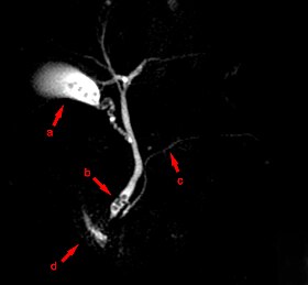
자기공명 담관 조영술(MRCP)은 의료 영상 기술입니다.자기공명영상을 사용하여 담도와 췌관을 비침습적으로 시각화합니다.이 절차는 담석이 담낭을 둘러싼 도관에 박혀 있는지 여부를 판단하기 위해 사용될 수 있다.
사용하다
MRCP는 선택한 조사로 내시경 역행 담관 조영술(ERCP)을 서서히 대체하고 있다.MRCP는 담도계, 췌장관을 진단하고 주변의 고형 장기에 접근하는데 매우 정확합니다.MRCP가 제공하는 몇 가지 장점은 ERCP(30분)에 비해 비침습적 특성, 비용, 검사 시간, 필요한 직원 수, 이온화 [1][2][3][4]방사선이 필요하지 않다는 것입니다.
MRCP는 담석 진단에 사용된다.그것은 또한 [5]매우 안정적으로 담즙낭종을 진단할 수 있다.담도계에 관한 정보를 제공하는 것 외에 MRCP는 주변의 고형 장기 및 혈관에 관한 정보를 제공하므로 췌장암 절제 계획 및 간경화나 담관암 [5]등의 원발성 경화성 담관염 합병증을 찾는 데 유용하다.
기술.
환자는 위장관 내 액체를 [1]최소한으로 유지하면서 담도계에 액체가 최대한 확장되도록 하기 위해 최소 4시간 동안 금식할 필요가 있다.그러나 스캔 [1]전에 투명한 액체와 정기적인 약물치료가 허용됩니다.파인애플 주스,[1] 대추 시럽, 페루목실, 아사이 주스 및 물과 같은 음의 구강 대비는 T2 신호 강도를 감소시켜 위 및 십이지장으로부터의 신호가 담도계 [6]신호와 간섭하는 것을 최소화하는데 유용하다.
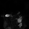
MRCP는 T2 가중치가 높은 MRI 펄스 [3][7]시퀀스를 사용합니다.이러한 시퀀스는 담낭, 담관 및 췌관 내의 정전기 또는 느린 유동액에서 높은 신호를 나타내며, 주변 조직의 신호는 낮습니다.세크레틴은 환자에게도 투여되어 도관 준수를 증가시켜 이미지를 더 쉽게 [3]만들 수 있습니다.
역사
그것은 [8]월너에 의해 1991년에 소개되었다.
기타 이미지
「 」를 참조해 주세요.
레퍼런스
- ^ a b c d Mandarano G, Sim J (October 2008). "The diagnostic MRCP examination: overcoming technical challenges to ensure clinical success". Biomedical Imaging and Intervention Journal. 4 (4): e28. doi:10.2349/biij.4.4.e28. PMC 3097748. PMID 21611015.
- ^ Prasad, SR; D. Sahani; S. Saini (November 2001). "Clinical applications of magnetic resonance cholangiopancreatography". Journal of Clinical Gastroenterology. 33 (5): 362–6. doi:10.1097/00004836-200111000-00004. PMID 11606850.
- ^ a b c Stevens, Tyler; Freeman, Martin L. (2019-01-01), Chandrasekhara, Vinay; Elmunzer, B. Joseph; Khashab, Mouen A.; Muthusamy, V. Raman (eds.), "57 - Recurrent Acute Pancreatitis", Clinical Gastrointestinal Endoscopy (Third Edition), Philadelphia: Elsevier, pp. 661–673.e3, doi:10.1016/b978-0-323-41509-5.00057-8, ISBN 978-0-323-41509-5, retrieved 2021-01-28
- ^ Hekimoglu K, Ustundag Y, Dusak A, et al. (August 2008). "MRCP vs. ERCP in the evaluation of biliary pathologies: review of current literature". Journal of Digestive Diseases. 9 (3): 162–9. doi:10.1111/j.1751-2980.2008.00339.x. PMID 18956595.
- ^ a b Fulcher, Ann S.; Turner, Mary Ann (2008-01-01), Gore, Richard M.; Levine, Marc S. (eds.), "chapter 77 - Magnetic Resonance Cholangiopancreatography", Textbook of Gastrointestinal Radiology (Third Edition), Philadelphia: W.B. Saunders, pp. 1383–1398, doi:10.1016/b978-1-4160-2332-6.50082-8, ISBN 978-1-4160-2332-6, retrieved 2021-01-28
- ^ Al-Atia, Mohassad. "Can oral contrast enhance image quality at MRCP? - A literature review" (PDF). Örebro University, Sweden. Part of Medicine, advanced level, Degree project. Archived from the original (PDF) on 12 June 2020. Retrieved 7 May 2022.
- ^ Griffin, Nyree; Charles-Edwards, Geoff; Grant, Lee Alexander (2011-09-28). "Magnetic resonance cholangiopancreatography: the ABC of MRCP". Insights into Imaging. 3 (1): 11–21. doi:10.1007/s13244-011-0129-9. ISSN 1869-4101. PMC 3292642. PMID 22695995.
- ^ Albert L. Baert (13 February 2008). Encyclopedia of Diagnostic Imaging. Springer. pp. 123–. ISBN 978-3-540-35278-5. Retrieved 3 July 2011.


