칸토넨시스오스트롱길루스
Angiostrongylus cantonensis| 칸토넨시스오스트롱길루스 | |
|---|---|
 | |
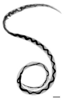
| 이발소 기둥 모양의 성충(지렁이의 안쪽 끝은 꼭대기까지).축척 막대는 1mm입니다. | |
| 과학적 분류 | |
| 왕국: | 애니멀리아 |
| 문: | 선충류 |
| 클래스: | 크로마도레아목 |
| 주문: | 라브디티다 |
| 패밀리: | 안지오스트롱길루스과 |
| 속: | 안지오스트롱길루스 |
| 종류: | 카토넨시스 |
| 이항명 | |
| 칸토넨시스오스트롱길루스 (Chen, 1935년)[1] | |
Angiostrongylus cantonensis는 동남아시아와 태평양 [2]유역에서 호산성 뇌수막염의 가장 흔한 원인인 Angiostrongyliais를 일으키는 기생 선충이다.선충은 일반적으로 쥐의 폐동맥에 존재하며, 쥐 [3]폐충이라는 별칭을 가지고 있다.달팽이는 애벌레가 감염될 때까지 자라는 주요 중간 숙주입니다.
인간은 이 회충의 부수적인 숙주로 날것이나 덜 익은 달팽이 또는 다른 벡터 또는 오염된 물과 [4]야채에 있는 애벌레를 섭취함으로써 감염될 수 있다.애벌레는 혈액을 통해 중추신경계로 운반되는데, 이들은 사망 또는 영구적인 뇌와 신경 [5]손상을 초래할 수 있는 심각한 병인 호산구 수막염의 가장 흔한 원인입니다.세계화가 질병의 [6][7]지리적 확산에 기여함에 따라, 혈관골격결핍증은 공중 보건의 중요성이 증가하는 감염이다.
역사
1935년 중국의 저명한 기생충학자 신타오첸(1904~1977)이 광둥 쥐 [1]표본을 조사한 결과 선충인 안지오스트롱길루스 광둥균이 1944년 노무라와 림에 의해 호산구성 뇌수막염 환자의 뇌척수액에서 확인됐다.그들은 환자가 먹은 날음식이 쥐에 의해 오염되었을 수 있다고 언급했다.1955년, 맥커래스와 샌더스는 달팽이와 민달팽이를 중간 숙주로 정의하고,[citation needed] 쥐의 혈액, 뇌, 폐를 통한 전염 경로를 주목하면서 쥐의 웜의 라이프사이클을 확인했습니다.
감염제

A. cantonensis는 선충문, 강직목, 상과 전이목의 곤충이다.선충은 단단한 외부 큐티클, 분할되지 않은 신체, 완전히 발달된 위장관을 특징으로 하는 회충이다.스트롱실리다목은 구충과 폐충을 포함한다.전이성 지렁이는 최종 [9]숙주의 폐에 존재하는 길이 [8]2cm의 가늘고 실 같은 벌레로 특징지어진다.Angiostrongylus costaricensis는 중앙아메리카와 남아메리카에서 장내 혈관조영증을 일으키는 밀접한 관련이 있는 벌레이다.
역학 및 병리발생
제2차 세계 대전 이후, A. cantonensis는 호주, 멜라네시아, 미크로네시아, 폴리네시아를 포함한 동남아시아와 서태평양 섬 전역에 퍼졌다.뉴칼레도니아, 필리핀, 라로통가, 사이판, 수마트라, 대만, 타히티에서 곧 환자가 보고되었다.1960년대에는 캄보디아, 괌, 하와이, 자바, 태국, 사라왁, 베트남, 바누아투 [10]등의 지역에서 더 많은 사례가 보고되었다.
1961년, 인간의 호산구 수막염에 대한 역학 연구가 로젠, 라이그레트, 보리스에 의해 이루어졌는데, 그는 이러한 감염을 일으키는 기생충이 물고기에 의해 옮겨진다는 가설을 세웠다.하지만, Alicata는 하와이에서 많은 수의 사람들에 의해 날 생선이 뚜렷한 결과 없이 소비되었고, 뇌수막염 증상을 보이는 환자들은 증상을 보이기 전에 몇 주 동안 날달팽이나 새우를 먹은 전력이 있다고 지적했다.감염된 뇌의 역학 및 부검과 함께, 이 관찰은 동남아시아와 태평양 [11]제도에서 호산성 뇌수막염의 대부분의 원인으로 인간에 대한 A. cantonensis 감염을 확인했다.
그 이후 미국령 사모아, 호주, 홍콩, 봄베이, 피지, 하와이, 혼슈, 인도, 규슈, 뉴브리튼, 오키나와, 류큐 제도, 서사모아, 그리고 가장 최근의 중국 본토에서 A. cantonensis 감염 사례가 나타났다.쥐 숙주에서 이 기생충의 다른 산발적인 발생은 쿠바, 이집트, 루이지애나, 마다가스카르, 나이지리아,[10] 푸에르토리코에서 보고되었다.
2013년 미국 플로리다에서 A. cantonensis가 발견되었으며, 그 범위와 유병률이 [12]확대되고 있다.2018년 [13]하와이를 방문한 뉴요커에게서 사례가 발견됐다.
최근 몇 [when?]년 동안, 기생충은 현대의 음식 소비 경향과 식품의 세계적인 운송으로 인해 놀라운 속도로 증식하고 있는 것으로 나타났다.과학자들은 A. cantonensis의 역학, 엄격한 식품 안전 정책, 그리고 중간 숙주 역할을 하는 달팽이와 민달팽이, 또는 물고기, 개구리, 또는 담수 같은 파라틴 숙주 역할을 하는 것과 같은 기생충에 의해 일반적으로 감염된 제품을 적절하게 소비하는 방법에 대한 지식의 증가를 요구하고 있다.새우.[14][15][16] 달팽이, 민달팽이 등 중간 숙주의 점액 배설물이나 최종 숙주 역할을 하는 쥐의 배설물에 의해 오염될 수 있는 식품을 섭취하는 것은 A. cantonensis의 [17]감염으로 이어질 수 있다.사람에게서 A. 광동성 감염의 가장 일반적인 경로는 [18]유충의 중간 숙주 또는 구충 숙주를 섭취하는 것이다.씻지 않은 과일과 야채, 특히 로메인 상추는 달팽이와 민달팽이 점액에 오염되거나 이러한 중간 숙주와 파라틴 숙주의 우발적인 섭취를 초래할 수 있습니다.이들 품목은 A. cantonensis 유충 또는 유충이 포함된 [19]숙주의 우발적인 섭취를 방지하기 위해 적절히 세척 및 취급할 필요가 있다.A. 광동 발생을 예방하는 가장 좋은 메커니즘은 달팽이와 민달팽이 개체수의 적극적인 통제, 적절한 음식 취급 기술과 함께 물고기, 민물 새우, 개구리, 연체동물, 달팽이와 같은 중간 및 구식 숙주의 적절한 조리 방법을 [20]도입하는 것입니다.설사에 대한 일반적인 예방법은 광동성 간염 [21]예방에 매우 효과적이다.왜 그것이 인간의 뇌를 목표로 하는지에 대해서는 많이 알려져 있지 않지만, 최근 화학적으로 유도되는 화학작용이 관련되어 있다.아세틸콜린은 니코틴성 아세틸콜린 [22]수용체를 통해 이 웜의 운동성을 증가시키는 것으로 이전에 보고되었다.A. 광동성 [23]감염에 따른 뇌 감염을 방지하기 위해 항콜린제 약물을 사용하여 화학적으로 유도되는 화학작용을 검증하기 위해 동물 모델의 실험 분석이 필요하다.
호스트
A. cantonensis의 유충의 중간 숙주는 다음과 같다.
- 육지 달팽이:Jamaica,[24][25]Pleurodonte sp에서 자메이카, Sagda sp에서 자메이카, Poteria sp에서 Jamaica,[25]Achatina fulica,[26][27][28][29]사쓰마 mercatoria, Acusta despecta,[28][29]Bradybaena brevispira,[26]Bradybaena에서Thelidomus aspera... Bradybaena ravida, 알았다고요에서 Bradybaena similaris, Plectotropis appanata[26]과 Parmarion martensi circulus[28].Inawa[28]과 Hawaii,[30]Camaena cicatricosa, Trichoch.Loritis ruopila, Trichloritis hungperfordianus, Cyclophorus spp.[29]
- 민물 달팽이 : Pila spp.,[27] Pomacea canaliculata,[26][27] Cipangopaludina chinensis, Bellamya aeruginosa, Bellamya quadrata[26]
- 민달팽이: 리맥스 막시무스,[31] 리맥스 플라버스[26] 데로케라스 레이브,[26][32] 데로케라스 레티큘라툼,[32] 라비카울리스 알테,[26][28][32] 사라시눌라 플레베아,[32] 바기눌루스 유시즈,[26][28] 리만니아 발렌티아나스, 피오로마이커스 빌리나투스, 마크로클라미사나, 메기마티쿠라툼 및 아마 다른[26] 민달팽이슬라툼의 종
A. cantonensis의 최종 숙주는 야생 설치류, 특히 갈색 쥐(Rattus norvegicus)와 검은 쥐(Rattus rattus)[24]를 포함한다.
A. cantonensis의 파라텐 숙주는 육지 편형동물 Platydemus manokwari와 양서류인 Bufo Asitiatus, Rana catesbeiana, Racophorus leucomystax 및 [28]Rana limnocaris를 포함한다.
2004년에는 노란꼬리검은코카투(Calyptorhynchus funereus)와 신경증상을 앓고 있는 자유생활하는 황갈색 개구리입(Podargus strigoides) 2마리가 기생충을 가진 것으로 나타났다.그들은 [33]유기체를 위해 발견된 첫 번째 조류 숙주였다.2018년 마요르카에서 급성 신경 질환의 징후가 있는 두 마리의 북아프리카 고슴도치가 뇌 속에 A. cantonensis가 있고, 그 중 한 마리는 육중한 [34]암컷이 있는 것으로 밝혀졌다.그것은 앤지오스트롱길루스의 [34]숙주로서 고슴도치에 대한 첫 보고였다.
하와이 보건부는 민물 오피히가 새우, 개구리, 물 모니터 [35]도마뱀과 같은 다른 수생 생물뿐만 아니라 기생충을 옮길 수 있다고 말한다.가정용 애완동물은 A. cantonensis를 운반하는 동물과 아직 잘 연구되지 않은 채 상호작용을 할 수 있다.고양이들은 고양이 폐충을 쥐와 달팽이의 [36]상호작용으로 옮기고 퍼뜨리는 것으로 알려져 있다.
인간 혈관오스트론지루시스 병원성
인간 중추신경계의 신경조직에 기생하는 벌레의 존재는 합병증을 일으킨다.다음의 모든 것에 의해,[citation needed] CNS 가 파손됩니다.
- 벌레의 움직임으로 인한 신경 조직에 직접적인 기계적 손상
- 질소 폐기물 등 유독 부산물
- 죽은 기생충과 살아있는 기생충에 의해 방출되는 항원
호산구성 수막염
비록 Angiostrongylus가 CNS에 침입함으로써 야기되는 임상적 질병은 일반적으로 "친환경성 수막염"이라고 불리고 있지만, 실제 병태생리학은 뇌수막, 또는 뇌의 표피 내벽에 침입하는 뇌수막염의 것이다.뇌수막의 내벽을 통한 초기 침입은 수막의 전형적인 염증과 두통, 뻣뻣한 목, 그리고 종종 발열의 전형적인 뇌수막염을 일으킬 수 있습니다.기생충은 이후 뇌 조직 깊숙이 침투하여 뇌 실질의 어디에 이동하느냐에 따라 특정한 국소 신경학적 증상을 일으킨다.초기 손상은 벌레의 물리적 이동에 의해 발생하며, 2차 손상은 죽은 벌레와 죽어가는 벌레의 존재에 대한 염증 반응에 의해 발생한다.이 염증은 단기적으로는 마비, 방광 기능 장애, 시각 장애, 혼수상태에 이를 수 있으며 장기적으로는 영구 신경 손상, 정신 지체, 신경 손상, 영구 뇌 손상 [37]또는 사망에 이를 수 있습니다.
호산구성 뇌수막염은 일반적으로 뇌척수액(CSF)의 호산구 수의 증가에 의해 정의된다.대부분의 경우 호산구 수치는 CSF에서 μl당 10개 이상의 호산구까지 상승하여 총 CSF 백혈구([38]백혈구) 카운트의 최소 10%를 차지한다.CSF의 화학적 분석은 일반적으로 단백질 수치, 정상 포도당 수치, 음성 세균 배양과 함께 "아제틱 수막염"의 발견과 유사합니다.CSF 분석에서 포도당의 유의한 감소는 심각한 뇌수막염의 지표이며 의학적 결과가 좋지 않음을 나타낼 수 있다.질병 과정의 초기 CSF 분석에서는 때때로 호산구 증가가 나타나지 않을 수 있지만, 후속 CSF 분석에서 호산구 증가가 있을 뿐이다.호산구 수막염은 지렁이가 [citation needed]CNS로 이동함에 따라 질병이 진화하기 때문에 전형적인 증상을 가진 사람의 혈관오스트롱길루스 감염을 진단하기 위한 유일한 기준으로 사용할 때 주의해야 한다.
호산구는 과립구 세포주의 특화된 백혈구로 세포질에 과립을 포함하고 있다.이 과립들은 기생충에 독성이 있는 단백질을 포함하고 있다.이 과립들이 분해되거나 분해되면, A. cantonensis와 같은 기생충과 싸우는 화학물질이 방출된다.몸 전체에 있는 호산구는 몸에 A. cantonensis와 같은 기생충이 들끓을 때 케모카인에 의해 염증 부위로 유도된다.일단 염증이 발생한 부위에 2형 사이토카인이 호산구와 소통하는 도우미 T세포에서 방출되어 활성화 신호를 보낸다.일단 활성화되면, 호산구는 외국 [citation needed]기생충과의 싸움에서 독성 단백질을 방출하면서 탈과립 과정을 시작할 수 있다.
임상적 징후 및 증상
그룹 사례 연구에 따르면, 가벼운 호산구 수막염에서 가장 흔한 증상은 두통, 광공포증 또는 시각 장애(92%), 목 경직(83%), 피로(83%), 과민증(75%), 구토(67%), 그리고 공포증(50%)[39][21]이다.잠복기는 보통 3주이지만 3-36일[10], 심지어 80일이 [40]될 수도 있다.
경미하고 심각한 호산구 수막염의 가능한 임상적 징후와 증상은 다음과 같다.
- 열은 종종 경미하거나 결석하지만, 고열의 존재는 심각한 [39]질병을 암시한다.
- 두통은 전두엽이나 후두엽의 [10]복두엽의 특징인 진행성이고 [39]심각하다.
- 수막 - 목이[39] 뻣뻣함
- 광공포증 - 빛에 대한[39] 민감성
- 근육 쇠약과 피로[10][40]
- 구토 유무에[10][39] 관계없이 메스꺼움
- 피부가 따끔거림, 따끔거림 또는 마비되는 증상은 몇 주 또는[10] 몇 달 동안 지속될 수 있습니다.
- 과민감 - 접촉에 대한 심한 민감성. 몇 주 또는[39] 몇 달 동안 지속될 수 있습니다.
- 방사상염 - 피부의 특정[39] 부위를 따라 조사되는 통증
- 방광기능부전증[41]
- 변비[41]
- 브루진스키 기호[40]
- 현기증
- 실명
- 한 부위에 국한된 마비(예: 안구외근육 마비 및 안면마비[40])
- 전신마비(완화증)[10]는 발에서 시작하여 몸 전체에 영향을 미치기 위해 위쪽으로 진행되는 경우가 많다.
- 혼수상태[39]
- 죽음[39]
치료
Angiostrongylus병의 심각도와 임상 과정은 3단계 [42]유충의 섭취 부하에 따라 크게 달라지며, 경우에 따라 큰 차이를 만들어 임상시험을 설계하기 어렵고 치료의 효과를 식별하기 어렵다.진통제와 진정제를 포함한 전형적인 보수적인 의료 관리는 두통과 과민증을 최소한 완화시켜 줍니다.3~7일 간격으로 뇌척수액을 제거하는 것은 두개골 내 압력을 유의하게 감소시키는 것으로 입증된 유일한 [43]방법이며 두통의 증상 치료에 사용될 수 있다.이 프로세스는 개선이 [38]나타날 때까지 반복할 수 있습니다.프레드니솔론이나[44] 덱사메타손을[45] 이용한 코르티코스테로이드 요법이 A. 칸토넨시스 [46][47]감염과 관련된 CNS 증상 치료에 도움이 된다는 적당한 품질의 증거가 증가하고 있다.초기의 연구antihelminthic 요원들을 가지고 치료되지 않았는지 또는 알벤다졸은 illness,[48][39]의 임상 코스 태국과 최근 중국에서 다양한 연구 개선에 효과적인thiobendazole 같은(마약 parasite-killing)이 glucocorticoids과 antihelminthics의 결합 durat을 감소시켜 안전한 것을 보여 준다.의 이온두통과 상당한 [45][44][49][50]두통을 가진 환자의 수.A. cantonensis 감염 관리를 위한 항헬름제의 첨가는 [43]유충의 동시 사망을 통해 과도한 양의 항원을 방출함으로써 신경학적 위기를 촉발시킬 이론적인 위험이 있지만, 임상 [51][45][50][44]환경에서 이러한 현상이 존재하는 것으로 밝혀진 연구는 없다.또한, 기생충이 CNS 밖으로 이동하기 전에 죽이는 데 실패하면 유충이 이동함으로써 기계적 손상의 위험이 높아집니다.알벤다졸과 프레드니솔론을 사용한 병용요법은 가벼운 [52]경우 프레드니솔론만을 사용한 치료에 비해 큰 이점이 없지만 항헬미네틱스 치료법은 확실히 안전하며 영구장애 또는 [37]사망위험이 있는 기생충 부하가 높은 환자에게 상당한 이점이 있을 수 있다.
진단.
A. cantonensis 감염으로 인한 질병의 진단은 종종 어렵고, 일반적으로 감염된 숙주의 섭취 가능 이력과 질병의 전형적인 특징의 존재에 크게 의존한다.호산구성 뇌수막염이 확인되었을 때 추정 진단이 특히 강하다.호산구성 수막염의 진단은 높은 두개골 압력과 호산구 수의 증가를 통해 도달할 수 있다.호산구성 수막염의 원인과 광동성 수막염의 존재에 대한 진단은 현저하게 더 어렵다.A. cantonensis 벌레 또는 유충을 찾기 위해 척추 탭 또는 CSF 샘플을 채취해야 합니다.A. cantonensis는 감염된 사람의 절반 이상에서 CSF에서 검출되지 않는다.현재 A. cantonensis와 관련된 특정 항원을 검출하는 방법도 신뢰할 수 없다.이에 따라 면역-PCR과 같은 항원-항체 반응을 검출하기 위한 대체 접근법이 모색되고 있다.[53]신속한 닷블롯 ELISA 테스트는 A. cantonensis의 [54]신속하고 효과적이며 경제적인 현장 진단에도 사용할 수 있습니다.
레퍼런스
- ^ a b Chen HT (1935). "Un nouveau nématode pulmonaire, Pulmonema cantonensis, n. g., n. sp" (PDF). Annales de Parasitologie Humaine et Comparée. 13 (4): 312–317. doi:10.1051/parasite/1935134312.

- ^ Baheti NN, Sreedharan M, Krishnamoorthy T, Nair MD, Radhakrishnan K (March 2008). "Neurological picture. Eosinophilic meningitis and an ocular worm in a patient from Kerala, south India". Journal of Neurology, Neurosurgery, and Psychiatry. 79 (3): 271. doi:10.1136/jnnp.2007.122093. PMID 18281446. S2CID 207001013.
- ^ Sheikh K (July 12, 2019). "In Hawaii, Rat Lungworm Disease Infects People but Eludes Researchers". The New York Times. Retrieved 13 July 2019.
- ^ Magill AJ, Strickland GT, Maguire JH, Ryan ET, Solomon T (2012). Hunter's tropical medicine and emerging infectious disease (9th ed.). London: Elsevier Health Sciences. ISBN 9781455740437. OCLC 861539914.
- ^ Li H, Xu F, Gu JB, Chen XG (October 2008). "A severe eosinophilic meningoencephalitis caused by infection of Angiostrongylus cantonensis". The American Journal of Tropical Medicine and Hygiene. 79 (4): 568–70. doi:10.4269/ajtmh.2008.79.568. PMID 18840746. S2CID 2094357.
- ^ Eamsobhana P (December 2014). "Eosinophilic meningitis caused by Angiostrongylus cantonensis--a neglected disease with escalating importance" (PDF). Tropical Biomedicine. 31 (4): 569–78. PMID 25776582. Archived from the original (PDF) on 2017-08-29.
- ^ Dard C, Tessier E, Nguyen D, Epelboin L, Harrois D, Swale C, et al. (2020). "First cases of Angiostrongylus cantonensis infection reported in Martinique, 2002-2017". Parasite. 27: 31. doi:10.1051/parasite/2020032. PMC 7216674. PMID 32394891.

- ^ Public Health Agency of Canada (2001-09-17). "Pathogen Safety Data Sheet: Infectious Substances – Angiostrongylus cantonensis - Canada.ca". www.phac-aspc.gc.ca. Archived from the original on 2017-07-14. Retrieved 2018-04-13.
- ^ "Helminth Taxonomy - Phylum Nematoda". Schistosomiasis Research Group. Archived from the original on April 13, 2009. Retrieved 26 February 2009.
- ^ a b c d e f g h "EOSINOPHILIC MENINGITIS" (PDF). louisiana.gov. 2018-03-09. Archived from the original (PDF) on 2010-06-15.
- ^ Alicata JE (June 1991). "The discovery of Angiostrongylus cantonensis as a cause of human eosinophilic meningitis". Parasitology Today. 7 (6): 151–3. doi:10.1016/0169-4758(91)90285-v. PMID 15463478.
- ^ Iwanowicz DD, Sanders LR, Schill WB, Xayavong MV, da Silva AJ, Qvarnstrom Y, Smith T (July 2015). "Spread of the Rat Lungworm (Angiostrongylus cantonensis) in Giant African Land Snails (Lissachatina fulica) in Florida, USA". Journal of Wildlife Diseases. 51 (3): 749–53. doi:10.7589/2014-06-160. PMID 25973628. S2CID 44951384.
- ^ "New York teen contracts rat lungworm during Hawaii vacation". NY Daily News. Archived from the original on 2018-04-13. Retrieved 2018-04-13.
- ^ Lv S, Zhang Y, Steinmann P, Zhou XN (January 2008). "Emerging angiostrongyliasis in Mainland China". Emerging Infectious Diseases. 14 (1): 161–4. doi:10.3201/eid1401.061529. PMC 2600175. PMID 18258099.
- ^ Alicata JE (1967). "Effect of Freezing and Boiling on the Infectivity of Third-Stage Larvae of Angiostrongylus cantonensis Present in Land Snails and Freshwater Prawns". The Journal of Parasitology. 53 (5): 1064–1066. doi:10.2307/3276839. JSTOR 3276839. PMID 6062057.
- ^ Ash LR (1968). "The Occurrence of Angiostrongylus cantonensis in Frogs of New Caledonia with Observations on Paratenic Hosts of Metastrongyles". The Journal of Parasitology. 54 (3): 432–436. doi:10.2307/3277060. JSTOR 3277060. PMID 5757723.
- ^ Richards CS, Merritt JW (April 1967). "Studies on Angiostrongylus cantonensis in molluscan intermediate hosts". The Journal of Parasitology. 53 (2): 382–8. doi:10.2307/3276595. JSTOR 3276595. PMID 6022396.
- ^ Campbell BG, Little MD (May 1988). "The finding of Angiostrongylus cantonensis in rats in New Orleans". The American Journal of Tropical Medicine and Hygiene. 38 (3): 568–73. CiteSeerX 10.1.1.890.7349. doi:10.4269/ajtmh.1988.38.568. PMID 3275136.
- ^ Yeung NW, Hayes KA, Cowie RH (June 2013). "Effects of washing produce contaminated with the snail and slug hosts of Angiostrongylus cantonensis with three common household solutions". Hawai'i Journal of Medicine & Public Health. 72 (6 Suppl 2): 83–6. PMC 3689494. PMID 23901391.
- ^ Cowie RH (June 2013). "Pathways for transmission of angiostrongyliasis and the risk of disease associated with them". Hawai'i Journal of Medicine & Public Health. 72 (6 Suppl 2): 70–4. PMC 3689478. PMID 23901388.
- ^ a b Slom TJ, Cortese MM, Gerber SI, Jones RC, Holtz TH, Lopez AS, Zambrano CH, Sufit RL, Sakolvaree Y, Chaicumpa W, Herwaldt BL, Johnson S (February 2002). "An outbreak of eosinophilic meningitis caused by Angiostrongylus cantonensis in travelers returning from the Caribbean" (PDF). The New England Journal of Medicine. 346 (9): 668–75. doi:10.1056/NEJMoa012462. PMID 11870244. Archived from the original (PDF) on 2017-08-09.
- ^ Mentz MB, Graeff-Teixeira C (July 2003). "Drug trials for treatment of human angiostrongyliasis" (PDF). Revista do Instituto de Medicina Tropical de Sao Paulo. 45 (4): 179–84. doi:10.1590/S0036-46652003000400001. PMID 14502343. Archived from the original (PDF) on 2018-10-30.
- ^ Baig AM (December 2017). "Is There a Cholinergic Survival Incentive for Neurotropic Parasites in the Brain?". ACS Chemical Neuroscience. 8 (12): 2574–2577. doi:10.1021/acschemneuro.7b00370. PMID 28985043.
- ^ a b Lindo JF, Waugh C, Hall J, Cunningham-Myrie C, Ashley D, Eberhard ML, Sullivan JJ, Bishop HS, Robinson DG, Holtz T, Robinson RD (March 2002). "Enzootic Angiostrongylus cantonensis in rats and snails after an outbreak of human eosinophilic meningitis, Jamaica". Emerging Infectious Diseases. 8 (3): 324–6. doi:10.3201/eid0803.010316. PMC 2732477. PMID 11927033.
- ^ a b Barth JD, Jansen H, Kromhout D, Reiber JH, Birkenhager JC, Arntzenius AC (November 1987). "Progression and regression of human coronary atherosclerosis. The role of lipoproteins, lipases and thyroid hormones in coronary lesion growth". Atherosclerosis. 68 (1–2): 51–8. doi:10.1016/0021-9150(87)90093-1. PMID 3689483.
- ^ a b c d e f g h i j Lv S, Zhang Y, Steinmann P, Zhou XN (January 2008). "Emerging angiostrongyliasis in Mainland China". Emerging Infectious Diseases. 14 (1): 161–4. doi:10.3201/eid1401.061529. PMC 2600175. PMID 18258099.
- ^ a b c Lv S, Zhang Y, Chen SR, Wang LB, Fang W, Chen F, Jiang JY, Li YL, Du ZW, Zhou XN (September 2009). Graeff-Teixeira C (ed.). "Human angiostrongyliasis outbreak in Dali, China". PLOS Neglected Tropical Diseases. 3 (9): e520. doi:10.1371/journal.pntd.0000520. PMC 2739427. PMID 19771154.

- ^ a b c d e f g Asato R, Taira K, Nakamura M, Kudaka J, Itokazu K, Kawanaka M (2004). "Changing Epidemiology of Angiostrongyliasis Cantonensis in Okinawa Prefecture, Japan" (PDF). Japanese Journal of Infectious Diseases. 57 (4): 184–186. PMID 15329455.
- ^ a b c "MEMORANDUM FOR 18 MDG/SGPM" (PDF). "Land snail infection rates for the human parasitic nematode, Angiostrongylus cantonensis (rat lung worm) with notes on snail and parasite biology and distribution on Kadena AB, Okinawa Japan. Consultative Letter, IERA-DO-BR-CL-2001-0049.". Department of the Air Force. 20 June 2001. Archived from the original (PDF) on 2011-08-11.
{{cite web}}: CS1 유지보수: 기타 (링크) - ^ Hollingsworth RG, Kaneta R, 설리반은 JJ, 비숍 HS, Qvarnstrom Y, 다 실바 AJ, 로빈슨은 댄 길버트(2007년).Parmarion 비교하라"분포.Martensi(유폐류:Helicarionidae), 새로운 Semi-Slug 해충 Hawai'i 섬에 건축물과 잠재력 인간 Angiostrongyliasis1"(PDF)에 대한 벡터다.퍼시픽 사이언스(원고 제출한)61(4):457–467. doi:10.2984[457:DOPCMP]2.0.CO;2.hdl:10125/22629.S2CID 14202324..
- ^ Senanayake SN, Pryor DS, Walker J, Konecny P (October 2003). "First report of human angiostrongyliasis acquired in Sydney" (PDF). The Medical Journal of Australia. 179 (8): 430–1. doi:10.5694/j.1326-5377.2003.tb05623.x. PMID 14558868. S2CID 27285797. Archived from the original (PDF) on 2019-05-31.
- ^ a b c d Högger CH (25 March 2003). "Antagonists of Slugs and Snails. A Bibliography of Sources and a List of Citations grouped according to Taxon of the Antagonists". Archived from the original on 2008-05-24.
- ^ Monks DJ, Carlisle MS, Carrigan M, Rose K, Spratt D, Gallagher A, Prociv P (2005). "Angiostrongylus cantonensis as a Cause of Cerebrospinal Disease in a Yellow-tailed Black Cockatoo (Calyptorhynchus funereus) and Two Tawny Frogmouths (Podargus strigoides)". Journal of Avian Medicine and Surgery. 19 (4): 289–293. doi:10.1647/2004-024.1. S2CID 86181695.
- ^ a b Paredes-Esquivel C, Sola J, Delgado-Serra S, Puig Riera M, Negre N, Miranda MÁ, Jurado-Rivera JA (August 2019). "Angiostrongylus cantonensis in North African hedgehogs as vertebrate hosts, Mallorca, Spain, October 2018". Euro Surveillance. 24 (33). doi:10.2807/1560-7917.ES.2019.24.33.1900489. PMC 6702795. PMID 31431209.
- ^ "Rat Lungworm Frequently Asked Questions the Daniel K. Inouye College of Pharmacy".
- ^ Colella V, Giannelli A, Brianti E, Ramos RA, Cantacessi C, Dantas-Torres F, Otranto D (August 2015). "Feline lungworms unlock a novel mode of parasite transmission". Scientific Reports. 5: 13105. Bibcode:2015NatSR...513105C. doi:10.1038/srep13105. PMC 4536521. PMID 26271902.
- ^ a b Wang QP, Lai DH, Zhu XQ, Chen XG, Lun ZR (October 2008). "Human angiostrongyliasis" (PDF). The Lancet. Infectious Diseases. 8 (10): 621–30. doi:10.1016/S1473-3099(08)70229-9. PMID 18922484. Archived from the original (PDF) on 2018-10-29.
- ^ a b 2006년 2월 28일 개정, 2011년 6월 1일에 접속.
- ^ a b c d e f g h i j k Murphy GS, Johnson S (June 2013). "Clinical aspects of eosinophilic meningitis and meningoencephalitis caused by Angiostrongylus cantonensis, the rat lungworm". Hawai'i Journal of Medicine & Public Health. 72 (6 Suppl 2): 35–40. PMC 3689484. PMID 23901382.
- ^ a b c d Tseng YT, Tsai HC, Sy CL, Lee SS, Wann SR, Wang YH, Chen JK, Wu KS, Chen YS (October 2011). "Clinical manifestations of eosinophilic meningitis caused by Angiostrongylus cantonensis: 18 years' experience in a medical center in southern Taiwan". Journal of Microbiology, Immunology, and Infection = Wei Mian Yu Gan Ran Za Zhi. 44 (5): 382–9. doi:10.1016/j.jmii.2011.01.034. PMID 21524976.

- ^ a b Maretić T, Perović M, Vince A, Lukas D, Dekumyoy P, Begovac J (June 2009). "Meningitis and radiculomyelitis caused by Angiostrongylus cantonensis". Emerging Infectious Diseases. 15 (6): 996–8. doi:10.3201/eid1506.081263. PMC 2727321. PMID 19523323.
- ^ Tsai HC, Liu YC, Kunin CM, Lee SS, Chen YS, Lin HH, et al. (August 2001). "Eosinophilic meningitis caused by Angiostrongylus cantonensis: report of 17 cases". The American Journal of Medicine. 111 (2): 109–14. doi:10.1016/S0002-9343(01)00766-5. PMID 11498063.
- ^ a b Slom TJ, Cortese MM, Gerber SI, Jones RC, Holtz TH, Lopez AS, et al. (February 2002). "An outbreak of eosinophilic meningitis caused by Angiostrongylus cantonensis in travelers returning from the Caribbean" (PDF). The New England Journal of Medicine. 346 (9): 668–75. doi:10.1056/nejmoa012462. PMID 11870244. Archived from the original (PDF) on 2017-08-09.
- ^ a b c Chotmongkol V (2006). "Treatment of eosinophilic meningitis with a combination of prednisolone and mebendazole". Am J Trop Med Hyg. 74 (6): 1122–1124. CiteSeerX 10.1.1.562.1854. doi:10.4269/ajtmh.2006.74.1122. PMID 16760531.
- ^ a b c Tsai TH, Liu YC, Wann SR, Lin WR, Lee SJ, Lin HH, et al. (March 2001). "An outbreak of meningitis caused by Angiostrongylus cantonensis in Kaohsiung". Journal of Microbiology, Immunology, and Infection = Wei Mian Yu Gan Ran Za Zhi. 34 (1): 50–6. PMID 11321128.
- ^ Chotmongkol V, Sawanyawisuth K, Thavornpitak Y (September 2000). "Corticosteroid treatment of eosinophilic meningitis". Clinical Infectious Diseases. 31 (3): 660–2. doi:10.1086/314036. PMID 11017811.

- ^ Thanaviratananich S, Thanaviratananich S, Ngamjarus C (February 2015). "Corticosteroids for parasitic eosinophilic meningitis". The Cochrane Database of Systematic Reviews (2): CD009088. doi:10.1002/14651858.CD009088.pub3. PMC 7111302. PMID 25687750.
- ^ Kliks MM, Kroenke K, Hardman JM (November 1982). "Eosinophilic radiculomyeloencephalitis: an angiostrongyliasis outbreak in American Samoa related to ingestion of Achatina fulica snails". The American Journal of Tropical Medicine and Hygiene. 31 (6): 1114–22. doi:10.4269/ajtmh.1982.31.1114. PMID 7149098.
- ^ Jitpimolmard S, Sawanyawisuth K, Morakote N, Vejjajiva A, Puntumetakul M, Sanchaisuriya K, et al. (May 2007). "Albendazole therapy for eosinophilic meningitis caused by Angiostrongylus cantonensis". Parasitology Research. 100 (6): 1293–6. doi:10.1007/s00436-006-0405-7. PMID 17177056. S2CID 6771800.
- ^ a b Chotmongkol V, Wongjitrat C, Sawadpanit K, Sawanyawisuth K (March 2004). "Treatment of eosinophilic meningitis with a combination of albendazole and corticosteroid" (PDF). The Southeast Asian Journal of Tropical Medicine and Public Health. 35 (1): 172–4. PMID 15272765. Archived from the original (PDF) on 2019-05-31.
- ^ Chotmongkol V, Sawanyawisuth K, Thavornpitak Y (September 2000). "Corticosteroid treatment of eosinophilic meningitis". Clinical Infectious Diseases. 31 (3): 660–2. doi:10.1086/314036. PMID 11017811.

- ^ Chotmongkol V, Kittimongkolma S, Niwattayakul K, Intapan PM, Thavornpitak Y (September 2009). "Comparison of prednisolone plus albendazole with prednisolone alone for treatment of patients with eosinophilic meningitis". The American Journal of Tropical Medicine and Hygiene. 81 (3): 443–5. doi:10.4269/ajtmh.2009.81.443. PMID 19706911.
- ^ Chye SM, Lin SR, Chen YL, Chung LY, Yen CM (January 2004). "Immuno-PCR for detection of antigen to Angiostrongylus cantonensis circulating fifth-stage worms". Clinical Chemistry. 50 (1): 51–7. doi:10.1373/clinchem.2003.020867. PMID 14709636.

- ^ Eamsobhana P, Yong HS (July 2009). "Immunological diagnosis of human angiostrongyliasis due to Angiostrongylus cantonensis (Nematoda: Angiostrongylidae)". International Journal of Infectious Diseases. 13 (4): 425–31. doi:10.1016/j.ijid.2008.09.021. PMID 19117782.

외부 링크
"Angiostrongylus". CDC.gov. Centers for Disease Control and Prevention. 2015-12-28. Retrieved 2017-04-04.
- "DPDx - Angiostrongyliasis". CDC. 2016-10-17. Retrieved 2017-04-04. 기생충 생물학, 이미지 갤러리, 검사실 진단 및 치료 정보 탭.
- 미국 국립의학도서관의 Angiostrongylus+cantonensis 의학 주제 제목(MeSH)
- Sydney Morning Herald의 인체 감염 사례, Angiostrongylus cantonensis 인체 감염 사례:삼키기 어렵다: 민달팽이를 먹는 것은 희귀병을 일으킨다.
- UF/IFAS Featuring Creatures 웹사이트에서 Angiostronglyus cantonensis.


