뇌 자기공명영상
Magnetic resonance imaging of the brain| 뇌와 뇌간 MRI | |
|---|---|
 뇌 MRI | |
| ICD-10-PCS | B030ZZZ |
| ICD-9-CM | 88.91 |
| OPS-301 코드 | 3-800, 3-820 |
뇌 자기공명영상(MRI)은 자기공명영상(MRI)을 이용해 전리방사선(X선)이나 방사능 추적기를 사용하지 않고도 뇌와 뇌시스템의 고품질 2차원 또는 3차원 영상을 제작한다.
역사
인간의 뇌에 대한 최초의 MR 이미지는 1978년 이안 로버트 영과 휴 클로우가 이끄는 EMI 연구소의 두 그룹의 연구원들에 의해 얻어졌다.[1] 1986년 찰스 L. 두물린과 하워드 R. General Electric은 Hart at General Electric이 MR 혈관조영술을 개발했고, Denis Le Bihan은 첫 이미지를 획득했고 후에 특허를 획득했다.[2][3] 1988년 아르노 빌링거와 동료들은 수용성 조영제가 관류 MRI에 사용될 수 있다는 것을 증명했다.[4] 1990년 AT&T 벨 연구소의 오가와 세이지 박사는 dHb로 산소가 고갈된 혈액이 자기장에 끌린다는 사실을 인지하고 기능자기공명영상(fMRI)의 기초가 되는 기술을 발견했다.[5]
1990년대 초, NIH에서 일하고 있는 피터 배서, 르 비한, 그리고 아론 필러, 솔직히 말해서 하우, 그리고 동료들은 확산 텐서 이미징(DTI)을 개발했다.[6][7][8][9] Joseph Hajnal, Young 및 Graeme Byder는 1992년 정상적인 백색 물질에서 높은 신호 영역을 입증하기 위해 FLAIR 펄스 시퀀스를 사용했다고 설명했다.[10] 같은 해, 존 데트르, 앨런 P. 코레츠키, 동료들은 동맥 스핀 라벨을 개발했다.[11] 1997년 위르겐 R. 레이첸바흐, E. 마크 해크, 워싱턴 대학의 동료들은 Susceptibility 가중치 영상물을 개발했다.[12]
인간 두뇌 3.0 T에서의 첫 연구는 1994년에 발표되었고,[13] 1998년 8 T.[14] 9.4 T(2006)[15]에서 최대 10.5 T(2019)까지 인간 두뇌에 대한 연구가 수행되었다.[16]
Paul Lauterbur와 Sir Peter Mansfield는 MRI와 관련된 발견으로 2003년 노벨 생리의학상을 받았다.

온전한 뇌(사후)의 가장 높은 공간 해상도 기록은 매사추세츠 종합병원에서 나온 100미크론이다. 데이터는 2019년 10월 30일 네이처에 게재됐다.[17][18]
적용들
머리의 컴퓨터 단층 촬영에 비해 뇌 MRI가 갖는 장점 중 하나는 조직 대비가 더 낫고 [19]뇌계를 볼 때 CT보다 유물이 적다는 것이다. MRI는 뇌하수체 영상에도 우수하다.[20] 그러나 초기 대뇌염을 식별하는 데는 덜 효과적일 수 있다.[21]
뇌진탕의 경우 진행성 신경학적 증상, 초점 신경학적 소견 또는 검사 시 두개골 골절 우려가 없는 한 MRI를 피해야 한다.[22] 뇌진탕 분석에서, 회복 중에 이루어지는 병태생리학적 메커니즘을 관찰하기 위해 분절 음이소트로피, 평균 확산성, 뇌혈류, 글로벌 연결성의 측정을 취할 수 있다.[23]
태아 뇌 분석에서 MRI는 초음파보다 교정에 대한 정보를 더 많이 제공한다.[24]
신경계 영상촬영에는 다음과 같은 다양한 영상촬영양식 또는 시퀀스를 사용할 수 있다.
- T-가중치1(T1W) 영상: 뇌척수액은 어둡다. T-가중1 영상은 정상적인 해부학적 구조를 시각화하는 데 유용하다.
- T-가중치2(T2W) 영상: CSF는 가볍지만 지방(따라서 백색 물질)은 T-가중치1 영상보다 어둡다. T가중치2 영상은 병리학 시각화에 유용하다.[25]
- 확산 가중 영상(DWI): DWI는 물 분자의 확산을 이용하여 MR 영상에서 대비를 생성한다.
- 양성자 밀도(PD) 영상: CSF는 양성자 수준이 비교적 높아 CSF가 밝게 보인다. 회백질은 백질보다 밝다.[26]

참고 항목
갤러리

T1 MRI의 뇌 부위
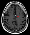
T1(CSF가 어둡다는 점 참조) 대비(팔록스의 뇌수종을 가리키는 화살표)
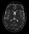
뇌의 정상 축 T2-가중 MR 영상
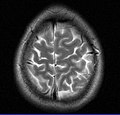
뇌 표면의 MRI 영상


| 위키미디어 커먼스는 뇌의 자기공명영상 관련 매체를 보유하고 있다. |
참조
- ^ Information, Reed Business (1978). "Britain's brains produce first NMR scans". New Scientist: 588.
- ^ "Blood-flow checker". Popular Science: 12. 1987.
- ^ Le Bihan D, Breton E (1987). "Method to Measure the Molecular Diffusion and/or Perfusion Parameters of Live Tissue". US Patent # 4,809,701.
- ^ Villringer A, Rosen BR, Belliveau JW, Ackerman JL, Lauffer RB, Buxton RB, Chao YS, Wedeen VJ, Brady TJ (February 1988). "Dynamic imaging with lanthanide chelates in normal brain: contrast due to magnetic susceptibility effects". Magnetic Resonance in Medicine. 6 (2): 164–74. doi:10.1002/mrm.1910060205. PMID 3367774. S2CID 41228095.
- ^ Faro SH, Mohamed FB (2010-01-15). Bold fMRI. a guide to functional imaging for neuroscientists. Springer. ISBN 978-1-4419-1328-9. Retrieved 10 June 2015.
- ^ Howe FA, Filler AG, Bell BA, Griffiths JR (December 1992). "Magnetic resonance neurography". Magnetic Resonance in Medicine. 28 (2): 328–38. doi:10.1002/mrm.1910280215. PMID 1461131. S2CID 36417513.
- ^ Filler AG, Howe FA, Hayes CE, Kliot M, Winn HR, Bell BA, Griffiths JR, Tsuruda JS (March 1993). "Magnetic resonance neurography". Lancet. 341 (8846): 659–61. doi:10.1016/0140-6736(93)90422-d. PMID 8095572. S2CID 24795253.
- ^ Filler A (October 2009). "Magnetic resonance neurography and diffusion tensor imaging: origins, history, and clinical impact of the first 50,000 cases with an assessment of efficacy and utility in a prospective 5000-patient study group". Neurosurgery. 65 (4 Suppl): A29-43. doi:10.1227/01.neu.0000351279.78110.00. PMC 2924821. PMID 19927075.
- ^ Basser PJ (2010). "Invention and Development of Diffusion Tensor MRI (DT-MRI or DTI) at the NIH". Diffusion MRI. Oxford University Press. pp. 730–740. doi:10.1093/med/9780195369779.003.0047. ISBN 9780195369779.
- ^ Hajnal JV, De Coene B, Lewis PD, Baudouin CJ, Cowan FM, Pennock JM, Young IR, Bydder GM (July 1992). "High signal regions in normal white matter shown by heavily T2-weighted CSF nulled IR sequences". Journal of Computer Assisted Tomography. 16 (4): 506–13. doi:10.1097/00004728-199207000-00002. PMID 1629405. S2CID 42727826.
- ^ Koretsky AP (August 2012). "Early development of arterial spin labeling to measure regional brain blood flow by MRI". NeuroImage. 62 (2): 602–7. doi:10.1016/j.neuroimage.2012.01.005. PMC 4199083. PMID 22245338.
- ^ Reichenbach JR, Venkatesan R, Schillinger DJ, Kido DK, Haacke EM (July 1997). "Small vessels in the human brain: MR venography with deoxyhemoglobin as an intrinsic contrast agent". Radiology. 204 (1): 272–7. doi:10.1148/radiology.204.1.9205259. PMID 9205259.
- ^ Mansfield P, Coxon R, Glover P (May 1994). "Echo-planar imaging of the brain at 3.0 T: first normal volunteer results". Journal of Computer Assisted Tomography. 18 (3): 339–43. doi:10.1097/00004728-199405000-00001. PMID 8188896. S2CID 20221062.
- ^ Robitaille PM, Abduljalil AM, Kangarlu A, Zhang X, Yu Y, Burgess R, Bair S, Noa P, Yang L, Zhu H, Palmer B, Jiang Z, Chakeres DM, Spigos D (October 1998). "Human magnetic resonance imaging at 8 T". NMR in Biomedicine. 11 (6): 263–5. doi:10.1002/(SICI)1099-1492(199810)11:6<263::AID-NBM549>3.0.CO;2-0. PMID 9802467.
- ^ Vaughan T; DelaBarre L; Snyder C; Tian J; Akgun C; Shrivastava D; Liu W; Olson C; Adriany G; et al. (December 2006). "9.4T human MRI: preliminary results". Magn Reson Med. 56 (6): 1274–82. doi:10.1002/mrm.21073. PMC 4406343. PMID 17075852.
- ^ Sadeghi‐Tarakameh, Alireza; DelaBarre, Lance; Lagore, Russell L.; Torrado‐Carvajal, Angel; Wu, Xiaoping; Grant, Andrea; Adriany, Gregor; Metzger, Gregory J.; Van de Moortele, Pierre‐Francois; Ugurbil, Kamil; Atalar, Ergin (2019-11-21). "In vivo human head MRI at 10.5T: A radiofrequency safety study and preliminary imaging results". Magnetic Resonance in Medicine. 84 (1): 484–496. doi:10.1002/mrm.28093. hdl:11693/53263. ISSN 0740-3194. PMC 7695227. PMID 31751499. S2CID 208226414.
- ^ "100-Hour-Long MRI of Human Brain Produces Most Detailed 3D Images Yet".
- ^ "Team publishes on highest resolution brain MRI scan".
- ^ Ebel K, Benz-Bohm G (1999). Differential diagnosis in pediatric radiology. Thieme. pp. 538–. ISBN 978-3-13-108131-5. Retrieved 18 July 2011.
- ^ Bradley WG, Brant-Zawadzki M, Cambray-Forker J (2001-01-15). MRI of the brain. Surendra Kumar. ISBN 978-0-7817-2568-2. Retrieved 24 July 2011.
- ^ Roos KL, Tunkel AR (2010). Bacterial infections of the central nervous system. Elsevier Health Sciences. pp. 69–. ISBN 978-0-444-52015-9. Retrieved 18 July 2011.
- ^ American Medical Society for Sports Medicine (24 April 2014), "Five Things Physicians and Patients Should Question", Choosing Wisely: an initiative of the ABIM Foundation, American Medical Society for Sports Medicine, retrieved 29 July 2014
- ^ Churchill Nathan W., Hutchison Michael G., Richards Doug, Leung General, Graham Simon J., Schweizer Tom A. (2017). "The first week after concussion: Blood flow, brain function and white matter microstructure". NeuroImage: Clinical. 14: 480–489. doi:10.1016/j.nicl.2017.02.015. PMC 5334547. PMID 28280686.
{{cite journal}}: CS1 maint : 복수이름 : 작성자 목록(링크) - ^ Garel C (2004). MRI of the fetal brain: normal development and cerebral pathologies. Springer. ISBN 978-3-540-40747-8. Retrieved 24 July 2011.
- ^ Butler P, Mitchell AW, Ellis H (2007-11-19). Applied Radiological Anatomy for Medical Students. Cambridge University Press. pp. 12–. ISBN 978-0-521-81939-8. Retrieved 18 July 2011.
- ^ Tofts, Paul (2005-09-01). Quantitative MRI of the Brain: Measuring Changes Caused by Disease. John Wiley and Sons. pp. 86–. ISBN 978-0-470-86949-9. Retrieved 18 July 2011.
- ^ Chowdhury R, Wilson I, Rofe C, Lloyd-Jones G (2010-04-19). Radiology at a Glance. John Wiley and Sons. pp. 95–. ISBN 978-1-4051-9220-0. Retrieved 18 July 2011.
- ^ Granacher RP (2007-12-20). Traumatic brain injury: methods for clinical and forensic neuropsychiatric assessment. CRC Press. pp. 247–. ISBN 978-0-8493-8138-6. Retrieved 18 July 2011.

