혈뇌장벽
Blood–brain barrier| 혈뇌장벽 | |
|---|---|
 BBB에서의 용질투과도 대 맥락막총 | |
| 세부 사항 | |
| 시스템. | 신경면역계 |
| 식별자 | |
| 머리글자 | BBB |
| MeSH | D001812 |
| 해부학 용어 [위키데이터 편집] | |
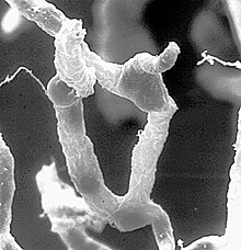
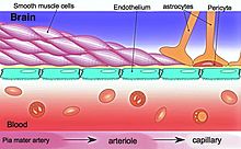
혈액-뇌 장벽(BBB)은 순환계와 중추신경계 사이에서 용질과 화학물질의 전달을 조절하는 매우 선택적인 내피 세포의 반투과성 경계로 혈액 내 유해하거나 원치 않는 물질로부터 뇌를 보호합니다.[1] 혈액-뇌 장벽은 모세혈관 벽의 내피 세포, 모세혈관을 덮고 있는 성상교세포 끝발, 그리고 모세혈관 기저막에 내장된 주변세포에 의해 형성됩니다.[2] 이 시스템은 수동적인 확산에 의한 일부 작은 분자의 통과뿐만 아니라 신경 기능에 중요한 각종 영양소, 이온, 유기 음이온, 포도당, 아미노산 등 거대 분자의 선택적이고 능동적인 운반을 가능하게 합니다.[3]
혈액-뇌 장벽은 병원체의 이동, 혈액 내 용질의 확산, 뇌척수액 내로의 큰 분자 또는 친수성 분자의 확산을 제한하는 동시에 소수성 분자(O2, CO2, 호르몬)와 작은 비극성 분자의 확산을 가능하게 합니다.[4][5] 장벽의 세포는 특정 수송 단백질을 사용하여 장벽을 가로질러 포도당과 같은 대사 생성물을 능동적으로 수송합니다.[6] 또한 이 장벽은 신호 분자, 항체 및 면역 세포와 같은 말초 면역 인자가 중추신경계로 전달되는 것을 제한하므로 뇌를 말초 면역 사건으로 인한 손상으로부터 절연시킵니다.[7]
뇌 신경 회로 내 감각 및 분비 통합에 참여하는 특수한 뇌 구조, 즉 심실 기관과 맥락막총은 대조적으로 투과성이 높은 모세혈관을 가지고 있습니다.[8]
구조.


BBB는 뇌 모세혈관의 내피 세포 사이의 단단한 접합부의 선택성으로 인해 용질의 통과가 제한됩니다.[1] 혈액과 뇌의 경계면에서, 내피 세포는 막횡단 단백질의 더 작은 소단위, 예를 들어, 클로딘(Claudin-5), 접합체 부착 분자(JAM-A)로 구성된 이러한 촘촘한 접합부에 의해 연속적으로 부착됩니다.[6] 이러한 각각의 타이트 접합 단백질은 타이트 접합 단백질 1(ZO1) 및 관련 단백질과 같은 스캐폴딩 단백질을 포함하는 다른 단백질 복합체에 의해 내피 세포막으로 안정화됩니다.[6]
BBB는 신체의 다른 곳에 있는 모세혈관의 내피세포보다 더 선택적으로 혈액에서 물질의 통과를 제한하는 내피세포로 구성되어 있습니다.[9] 성상교세포 발("glia limitans"라고도 함)이라고 불리는 성상교세포 세포 돌출부는 BBB의 내피 세포를 둘러싸고 있어 해당 세포에 생화학적 지원을 제공합니다.[10] BBB는 맥락막총의 맥락막 세포의 기능인 매우 유사한 혈액-뇌척수액 장벽과 그러한 장벽의 전체 영역의 일부로 간주될 수 있는 혈액-망막 장벽과 구별됩니다.[11]
인간 뇌의 모든 혈관이 BBB의 특성을 나타내는 것은 아닙니다. 여기에는 심실 기관, 제3, 제4심실의 지붕, 간뇌와 송과선의 지붕에 있는 송과선의 모세혈관이 포함됩니다. 송과선은 멜라토닌 호르몬을 "직접적으로" 분비하기 [12]때문에 멜라토닌은 혈액-뇌 장벽의 영향을 받지 않습니다.[13]
발전
BBB는 출생 시에 기능하는 것으로 보입니다. 수송체인 P-당단백질은 이미 배아 내피에 존재합니다.[14]
다양한 혈액 매개 용질의 뇌 흡수를 측정한 결과 신생아 내피 세포가 성인과 기능적으로 유사한 것으로 나타났으며,[15] 이는 선택적 BBB가 태어날 때 작동한다는 것을 나타냅니다.
마우스에서 발달 중 Claudin-5 손실은 치명적이며 BBB의 크기 선택적 이완을 초래합니다.[16]
기능.
혈액-뇌 장벽은 순환하는 병원체 및 기타 잠재적인 독성 물질로부터 뇌 조직을 보호하는 효과적인 역할을 합니다.[17] 따라서 뇌의 혈액 매개 감염은 거의 없습니다.[1] 발생하는 뇌의 감염은 종종 치료가 어렵습니다. 항체가 너무 커서 혈액-뇌 장벽을 통과할 수 없고, 특정 항생제만 통과할 수 있습니다.[18] 혈액-뇌척수액 장벽을 넘어 뇌로 들어갈 수 있는 뇌척수액에 직접 약물을 투여해야 하는 경우도 있습니다.[19][20]
뇌실외기관
심실외기(Circuitrical Organs, CVO)는 뇌의 제4심실 또는 제3심실에 인접하게 위치한 개별 구조물로, 혈액-뇌 장벽과 달리 투과성 내피세포가 밀집된 모세혈관층이 특징입니다.[21][22] 투과성 모세혈관이 높은 CVO 중에는 영역 후, 하부 기관지, 라미나 말단의 혈관 기관, 중간 신장, 송과선 및 뇌하수체의 3개 엽이 포함됩니다.[21][23]
감각적인 CVO(후유부, 하부정맥, 라미나 말단의 혈관 기관)의 투과성 모세혈관은 전신 혈액의 순환 신호를 신속하게 감지할 수 있게 하는 반면, 분비성 CVO(중간, 송과선, 뇌하수체엽)의 투과성 모세혈관은 뇌에서 파생된 신호를 순환 혈액으로 운반하는 것을 용이하게 합니다.[21][22] 결과적으로, CVO 투과성 모세혈관은 신경내분비 기능을 위한 양방향 혈액-뇌 의사소통의 지점입니다.[21][23][24]
투과성 특화구역
혈액-뇌 장벽의 "뒤"에 있는 뇌 조직과 특정 CVO에서 혈액 신호에 대해 "열린" 영역 사이의 경계 영역은 일반적인 뇌 모세혈관보다 누출이 많지만 CVO 모세혈관만큼 투과성이 없는 특수한 하이브리드 모세혈관을 포함합니다. 이러한 영역은 상복부(nucleus tractus solitarii, NTS)[25] 및 중앙부(median eminance, 시상하부(hypothalamic arcute nucleus)의 경계에 존재합니다.[24][26] 이 영역들은 NTS 및 아치형 핵과 같은 다양한 신경 회로와 관련된 뇌 구조가 혈액 신호를 수신한 다음 신경 출력으로 전달되는 빠른 통과 영역으로 기능하는 것으로 보입니다.[24][25] 중앙부 절제면과 시상하부 아치형 핵 사이에 공유되는 투과성 모세관 영역은 넓은 모세관 공간에 의해 증가되어 두 구조 사이에서 용질의 양방향 흐름을 촉진하고 중앙부 절제면이 분비 기관일 뿐만 아니라 감각 기관일 수 있음을 나타냅니다.[24][26]
치료연구
약물 표적으로
혈액-뇌 장벽은 뇌 모세혈관 내피에 의해 형성되며 대분자 신경치료제의 100%와 모든 소분자 약물의 98% 이상을 뇌에서 배제합니다.[1] 뇌의 특정 부위에 치료제를 전달하는 어려움을 극복하는 것은 대부분의 뇌 질환 치료에 큰 도전이 됩니다.[27][28] 신경 보호 역할에서 혈액-뇌 장벽은 잠재적으로 중요한 많은 진단 및 치료제가 뇌로 전달되는 것을 방해하는 기능을 합니다. 진단 및 치료에 효과적일 수 있는 치료 분자 및 항체는 임상적으로 효과적일 수 있는 적절한 양으로 BBB를 통과하지 않습니다.[27] BBB는 일부 약물이 뇌에 도달하는 데 장애물을 나타내므로 이 장벽을 극복하기 위해 BBB를 자연스럽게 통과할 수 있는 일부 펩티드가 약물 전달 시스템으로 널리 조사되었습니다.[29]
뇌에서 약물 표적화를 위한 메커니즘은 BBB를 "통하게" 또는 "뒤"하는 것을 포함합니다. BBB를 통해 단위 용량으로 뇌에 약물을 전달하는 방식은 삼투압 수단 또는 생화학적으로 브래디키닌과 같은 혈관 활성 물질을 사용하거나 [30]심지어 고강도 집속 초음파(HIFU)에 국부적으로 노출됨으로써 중단을 수반합니다.[31]
BBB를 통과하는 데 사용되는 다른 방법에는 포도당 및 아미노산 운반체와 같은 운반체 매개 수송체, 인슐린 또는 트랜스페린에 대한 수용체 매개 형질전환, p-당단백질과 같은 활성 유출 수송체의 차단을 포함한 내인성 수송 시스템의 사용이 수반될 수 있습니다.[27] 일부 연구에서는 트랜스페린 수용체와 같은 BBB 수송체를 표적으로 하는 벡터가 BBB를 가로질러 표적 부위로 운반되는 대신 모세혈관의 뇌 내피 세포에 계속 갇혀 있는 것으로 밝혀졌습니다.[27][32]
나노입자
나노기술은 BBB 전역의 약물 전달을 촉진할 수 있는 잠재력에 대해 예비 연구 중입니다.[27][33][34] 모세혈관 내피 세포 및 관련된 주변 세포는 종양에서 비정상적일 수 있으며 혈액-뇌 장벽이 뇌종양에서 항상 손상되지 않을 수 있습니다.[34] 성상세포와 같은 다른 요인은 나노입자를 이용한 치료에 대한 뇌종양의 내성에 기여할 수 있습니다.[35] 400달톤 미만의 지용성 분자는 지질 매개 수동 확산을 통해 BBB를 자유롭게 확산할 수 있습니다.[36]
상해 및 질병의 손상
알츠하이머병, 근위축성 측색 경화증, 뇌전증, 허혈성 뇌졸중, 뇌 외상에 대한 신경 영상 연구에서 알 수 있듯이, 혈액-뇌 장벽은 일부 신경 질환과 간 기능 부전과 같은 전신 질환에서 손상될 수 있습니다. 포도당 수송 장애 및 내피 변성과 같은 효과는 뇌 내 대사 기능 장애를 유발할 수 있으며, 전염증 인자에 대한 BBB의 투과성을 증가시켜 잠재적으로 항생제와 식세포가 BBB를 가로질러 이동할 수 있습니다.[1][27]
예측
실험적인 혈액-뇌 장벽 투과성과 물리화학적 특성을 연관시키려는 많은 시도가 있었습니다. 뇌-혈액 분포에 대한 최초의 QSAR 연구는 1988년에 수행되었으며, 이 연구는 많은 수의 H2 수용체 히스타민 작용제에 대한 쥐의 생체 내 값을 보고했습니다.[41] 혈액뇌장벽 투과성을 모델링하려는 최초의 논문들은 세 가지 특성, 즉 분자 부피, 친유성 및 수소 결합 가능성을 확인하여 혈액뇌장벽을 통과하는 수송에 크게 기여했습니다.[42] 수치 logBB 값(1058 분자)과 범주형 레이블(4956 BBB+ 및 2851 BBB-가 있는 7807 분자)의 두 데이터 세트가 2021년에 발표되었습니다.[43] 범주형 데이터 세트는 분자 지문,[45] MACCS166 키[46] 및 분자 기술자를 기반으로 4가지 다른 분류 모델을 선택하는[44] 데 2022년에 사용되었습니다.[47]
역사
1898년, 아서 비들과 R. 크라우스는 저농도의 "담즙염"이 동물의 혈류에 주입되었을 때 행동에 영향을 미치지 못하는 것을 관찰했습니다. 따라서, 이론적으로, 그들은 뇌로 들어가는 것에 실패했습니다.[48]
2년 후, 맥스 레반도프스키는 1900년에 "혈뇌 장벽"이라는 용어를 처음으로 만들어 냈을 것입니다.[49] 혈액-뇌 장벽이라는 용어는 레반도프스키의 것으로 추정되는 경우가 많아 이에 대한 논란이 있지만, 그의 논문에는 등장하지 않습니다. 이 용어의 창시자는 아마 리나 스턴이었을 것입니다.[50] 스턴은 러시아 과학자로 그녀의 연구를 러시아어와 프랑스어로 출판했습니다. 그녀의 출판물과 영어권 과학자들 사이의 언어 장벽 때문에, 이것은 그녀의 연구를 그 용어의 덜 알려진 기원으로 만들 수 있었습니다.
그 동안 세균학자 Paul Ehrlich는 화학 염료를 사용하여 미세한 생물학적 구조를 가시화하기 위해 많은 현미경 연구에서 사용되는 절차인 염색을 연구하고 있었습니다.[51] 에를리히가 이 염료들 중 일부(특히 당시 널리 사용되었던 아닐린 염료)를 주입했을 때, 그 염료는 뇌를 제외한 어떤 종류의 동물들의 모든 장기를 염색했습니다.[51] 그 당시, 에르리히는 염색의 부족을 단순히 뇌가 염료를 많이 감지하지 못하기 때문이라고 생각했습니다.[49]
그러나 1913년 이후의 실험에서 에드윈 골드만(Edwin Goldmann, Ehrlich의 학생 중 한 명)은 동물의 뇌척수액에 직접 염료를 주입했습니다. 그리고 나서 그는 뇌가 염색된 것을 발견했지만 나머지 신체는 염색되지 않아 둘 사이의 구획이 존재한다는 것을 보여주었습니다. 당시에는 뚜렷한 막을 찾을 수 없었기 때문에 혈관 자체가 장벽의 책임이 있다고 생각했습니다.
참고 항목
- 혈액-안구 장벽 – 국소 혈관과 눈의 대부분 부분 사이의 물리적 장벽
- 혈액-망막 장벽 – 특정 물질이 망막으로 들어오는 것을 막는 혈액-안구 장벽의 일부
- 혈액-타액 장벽 – 혈액 성분이 타액과 등으로 선택적으로 통과할 수 있는 반투과성 경계
- 혈액-척수 장벽 – 반투과성 해부학적 인터페이스
- 혈액-고환 장벽 – 혈관과 동물 고환의 정소 세뇨관 사이의 물리적 장벽
참고문헌
- ^ a b c d e f Daneman R, Prat A (January 2015). "The blood-brain barrier". Cold Spring Harbor Perspectives in Biology. 7 (1): a020412. doi:10.1101/cshperspect.a020412. PMC 4292164. PMID 25561720.
- ^ Ballabh P, Braun A, Nedergaard M (June 2004). "The blood-brain barrier: an overview: structure, regulation, and clinical implications". Neurobiology of Disease. 16 (1): 1–13. doi:10.1016/j.nbd.2003.12.016. PMID 15207256. S2CID 2202060.
- ^ Gupta S, Dhanda S, Sandhir R (2019). "Anatomy and physiology of blood-brain barrier". In Gao H, Gao X (eds.). Brain Targeted Drug Delivery System. Academic Press. pp. 7–31. doi:10.1016/b978-0-12-814001-7.00002-0. ISBN 978-0-12-814001-7. S2CID 91847478. Retrieved 2023-11-02.
- ^ Obermeier B, Daneman R, Ransohoff RM (December 2013). "Development, maintenance and disruption of the blood-brain barrier". Nature Medicine. 19 (12): 1584–96. doi:10.1038/nm.3407. PMC 4080800. PMID 24309662.
- ^ Kadry H, Noorani B, Cucullo L (November 2020). "A blood-brain barrier overview on structure, function, impairment, and biomarkers of integrity". Fluids Barriers CNS. 17 (1): 69. doi:10.1186/s12987-020-00230-3. PMC 7672931. PMID 33208141.
- ^ a b c Stamatovic SM, Keep RF, Andjelkovic AV (September 2008). "Brain endothelial cell-cell junctions: how to "open" the blood brain barrier". Current Neuropharmacology. 6 (3): 179–92. doi:10.2174/157015908785777210. PMC 2687937. PMID 19506719.
- ^ Muldoon LL, Alvarez JI, Begley DJ, Boado RJ, Del Zoppo GJ, Doolittle ND, et al. (January 2013). "Immunologic privilege in the central nervous system and the blood-brain barrier". Journal of Cerebral Blood Flow and Metabolism. 33 (1): 13–21. doi:10.1038/jcbfm.2012.153. PMC 3597357. PMID 23072749.
- ^ Kaur C, Ling EA (September 2017). "The circumventricular organs". Histology and Histopathology. 32 (9): 879–892. doi:10.14670/HH-11-881. PMID 28177105.
- ^ van Leeuwen LM, Evans RJ, Jim KK, Verboom T, Fang X, Bojarczuk A, et al. (February 2018). "A transgenic zebrafish model for the in vivo study of the blood and choroid plexus brain barriers using claudin 5". Biology Open. 7 (2): bio030494. doi:10.1242/bio.030494. PMC 5861362. PMID 29437557.
- ^ Abbott NJ, Rönnbäck L, Hansson E (January 2006). "Astrocyte-endothelial interactions at the blood-brain barrier". Nature Reviews. Neuroscience. 7 (1): 41–53. doi:10.1038/nrn1824. PMID 16371949. S2CID 205500476.
- ^ Hamilton RD, Foss AJ, Leach L (December 2007). "Establishment of a human in vitro model of the outer blood-retinal barrier". Journal of Anatomy. 211 (6): 707–16. doi:10.1111/j.1469-7580.2007.00812.x. PMC 2375847. PMID 17922819.

- ^ Pritchard TC, Alloway KD (1999). Medical Neuroscience (1st ed.). Fence Creek Publishing. pp. 76–77. ISBN 978-1-889325-29-3. OCLC 41086829.
- ^ Gilgun-Sherki Y, Melamed E, Offen D (June 2001). "Oxidative stress induced-neurodegenerative diseases: the need for antioxidants that penetrate the blood brain barrier". Neuropharmacology. 40 (8): 959–75. doi:10.1016/S0028-3908(01)00019-3. PMID 11406187. S2CID 15395925.
- ^ Tsai CE, Daood MJ, Lane RH, Hansen TW, Gruetzmacher EM, Watchko JF (January 2002). "P-glycoprotein expression in mouse brain increases with maturation". Biology of the Neonate. 81 (1): 58–64. doi:10.1159/000047185. PMID 11803178. S2CID 46815691.
- ^ Braun LD, Cornford EM, Oldendorf WH (January 1980). "Newborn rabbit blood-brain barrier is selectively permeable and differs substantially from the adult". Journal of Neurochemistry. 34 (1): 147–52. doi:10.1111/j.1471-4159.1980.tb04633.x. PMID 7452231. S2CID 21944159.
- ^ Nitta T, Hata M, Gotoh S, Seo Y, Sasaki H, Hashimoto N, et al. (May 2003). "Size-selective loosening of the blood-brain barrier in claudin-5-deficient mice". The Journal of Cell Biology. 161 (3): 653–60. doi:10.1083/jcb.200302070. PMC 2172943. PMID 12743111.
- ^ Abdullahi W, Tripathi D, Ronaldson PT (September 2018). "Blood-brain barrier dysfunction in ischemic stroke: targeting tight junctions and transporters for vascular protection". American Journal of Physiology. Cell Physiology. 315 (3): C343–C356. doi:10.1152/ajpcell.00095.2018. PMC 6171039. PMID 29949404.
- ^ Raza MW, Shad A, Pedler SJ, Karamat KA (March 2005). "Penetration and activity of antibiotics in brain abscess". Journal of the College of Physicians and Surgeons--Pakistan. 15 (3): 165–167. PMID 15808097.
- ^ Pardridge WM (January 2011). "Drug transport in brain via the cerebrospinal fluid". Fluids and Barriers of the CNS. 8 (1): 7. doi:10.1186/2045-8118-8-7. PMC 3042981. PMID 21349155.
- ^ Chen Y, Imai H, Ito A, Saito N (2013). "Novel modified method for injection into the cerebrospinal fluid via the cerebellomedullary cistern in mice". Acta Neurobiologiae Experimentalis. 73 (2): 304–311. PMID 23823990.
- ^ a b c d Gross PM, Weindl A (December 1987). "Peering through the windows of the brain". Journal of Cerebral Blood Flow and Metabolism. 7 (6): 663–72. doi:10.1038/jcbfm.1987.120. PMID 2891718.
- ^ a b Gross PM (1992). "Chapter 31: Circumventricular organ capillaries". Circumventricular Organs and Brain Fluid Environment - Molecular and Functional Aspects. Progress in Brain Research. Vol. 91. pp. 219–33. doi:10.1016/S0079-6123(08)62338-9. ISBN 9780444814197. PMID 1410407.
- ^ a b Miyata S (2015). "New aspects in fenestrated capillary and tissue dynamics in the sensory circumventricular organs of adult brains". Frontiers in Neuroscience. 9: 390. doi:10.3389/fnins.2015.00390. PMC 4621430. PMID 26578857.
- ^ a b c d Rodríguez EM, Blázquez JL, Guerra M (April 2010). "The design of barriers in the hypothalamus allows the median eminence and the arcuate nucleus to enjoy private milieus: the former opens to the portal blood and the latter to the cerebrospinal fluid". Peptides. 31 (4): 757–76. doi:10.1016/j.peptides.2010.01.003. PMID 20093161. S2CID 44760261.
- ^ a b Gross PM, Wall KM, Pang JJ, Shaver SW, Wainman DS (December 1990). "Microvascular specializations promoting rapid interstitial solute dispersion in nucleus tractus solitarius". The American Journal of Physiology. 259 (6 Pt 2): R1131-8. doi:10.1152/ajpregu.1990.259.6.R1131. PMID 2260724.
- ^ a b Shaver SW, Pang JJ, Wainman DS, Wall KM, Gross PM (March 1992). "Morphology and function of capillary networks in subregions of the rat tuber cinereum". Cell and Tissue Research. 267 (3): 437–48. doi:10.1007/BF00319366. PMID 1571958. S2CID 27789146.
- ^ a b c d e f g Sweeney MD, Sagare AP, Zlokovic BV (March 2018). "Blood-brain barrier breakdown in Alzheimer disease and other neurodegenerative disorders". Nature Reviews. Neurology. 14 (3): 133–150. doi:10.1038/nrneurol.2017.188. PMC 5829048. PMID 29377008.
- ^ Harilal S, Jose J, Parambi DG, Kumar R, Unnikrishnan MK, Uddin MS, et al. (July 2020). "Revisiting the blood-brain barrier: A hard nut to crack in the transportation of drug molecules". Brain Research Bulletin. 160: 121–140. doi:10.1016/j.brainresbull.2020.03.018. PMID 32315731. S2CID 215807970.
- ^ de Oliveira EC, da Costa KS, Taube PS, Lima AH, Junior CS (2022-03-25). "Biological Membrane-Penetrating Peptides: Computational Prediction and Applications". Frontiers in Cellular and Infection Microbiology. 12: 838259. doi:10.3389/fcimb.2022.838259. PMC 8992797. PMID 35402305.
- ^ Marcos-Contreras OA, Martinez de Lizarrondo S, Bardou I, Orset C, Pruvost M, Anfray A, et al. (November 2016). "Hyperfibrinolysis increases blood-brain barrier permeability by a plasmin- and bradykinin-dependent mechanism". Blood. 128 (20): 2423–2434. doi:10.1182/blood-2016-03-705384. PMID 27531677.
- ^ McDannold N, Vykhodtseva N, Hynynen K (May 2008). "Blood-brain barrier disruption induced by focused ultrasound and circulating preformed microbubbles appears to be characterized by the mechanical index". Ultrasound in Medicine & Biology. 34 (5): 834–40. doi:10.1016/j.ultrasmedbio.2007.10.016. PMC 2442477. PMID 18207311.
- ^ Wiley DT, Webster P, Gale A, Davis ME (May 2013). "Transcytosis and brain uptake of transferrin-containing nanoparticles by tuning avidity to transferrin receptor". Proceedings of the National Academy of Sciences of the United States of America. 110 (21): 8662–7. Bibcode:2013PNAS..110.8662W. doi:10.1073/pnas.1307152110. PMC 3666717. PMID 23650374.
- ^ Krol S, Macrez R, Docagne F, Defer G, Laurent S, Rahman M, et al. (March 2013). "Therapeutic benefits from nanoparticles: the potential significance of nanoscience in diseases with compromise to the blood brain barrier". Chemical Reviews. 113 (3): 1877–903. doi:10.1021/cr200472g. PMID 23157552.
- ^ a b Silva GA (December 2008). "Nanotechnology approaches to crossing the blood-brain barrier and drug delivery to the CNS". BMC Neuroscience. 9 (Suppl 3): S4. doi:10.1186/1471-2202-9-S3-S4. PMC 2604882. PMID 19091001.
- ^ Hashizume H, Baluk P, Morikawa S, McLean JW, Thurston G, Roberge S, et al. (April 2000). "Openings between defective endothelial cells explain tumor vessel leakiness". The American Journal of Pathology. 156 (4): 1363–80. doi:10.1016/S0002-9440(10)65006-7. PMC 1876882. PMID 10751361.
- ^ Souza RM, da Silva IC, Delgado AB, da Silva PH, Costa VR (2018). "Focused ultrasound and Alzheimer's disease A systematic review". Dementia & Neuropsychologia. 12 (4): 353–359. doi:10.1590/1980-57642018dn12-040003. PMC 6289486. PMID 30546844.
- ^ Abdullahi W, Tripathi D, Ronaldson PT (September 2018). "Blood-brain barrier dysfunction in ischemic stroke: targeting tight junctions and transporters for vascular protection". American Journal of Physiology. Cell Physiology. 315 (3): C343–C356. doi:10.1152/ajpcell.00095.2018. PMC 6171039. PMID 29949404.
- ^ Turner RJ, Sharp FR (2016-03-04). "Implications of MMP9 for Blood Brain Barrier Disruption and Hemorrhagic Transformation Following Ischemic Stroke". Frontiers in Cellular Neuroscience. 10: 56. doi:10.3389/fncel.2016.00056. PMC 4777722. PMID 26973468.
- ^ Mracsko E, Veltkamp R (2014-11-20). "Neuroinflammation after intracerebral hemorrhage". Frontiers in Cellular Neuroscience. 8: 388. doi:10.3389/fncel.2014.00388. PMC 4238323. PMID 25477782.
- ^ Alluri H, Wiggins-Dohlvik K, Davis ML, Huang JH, Tharakan B (October 2015). "Blood-brain barrier dysfunction following traumatic brain injury". Metabolic Brain Disease. 30 (5): 1093–1104. doi:10.1007/s11011-015-9651-7. PMID 25624154. S2CID 17688028.
- ^ Young RC, Mitchell RC, Brown TH, Ganellin CR, Griffiths R, Jones M, Rana KK, Saunders D, Smith IR (March 1988). "Development of a new physicochemical model for brain penetration and its application to the design of centrally acting H2 receptor histamine antagonists". Journal of Medicinal Chemistry. 31 (3): 656–671. doi:10.1021/jm00398a028. eISSN 1520-4804. ISSN 0022-2623. PMID 2894467.
- ^ Zhang L, Zhu H, Oprea TI, Golbraikh A, Tropsha A (14 June 2008). "QSAR Modeling of the Blood–Brain Barrier Permeability for Diverse Organic Compounds". Pharmaceutical Research. 25 (8): 1902–1914. doi:10.1007/s11095-008-9609-0. eISSN 1573-904X. ISSN 0724-8741. PMID 18553217. S2CID 22184045.
- ^ Meng F, Xi Y, Huang J, Ayers PW (2021-10-29). "A curated diverse molecular database of blood-brain barrier permeability with chemical descriptors". Scientific Data. Springer Science and Business Media LLC. 8 (1): 289. Bibcode:2021NatSD...8..289M. doi:10.1038/s41597-021-01069-5. ISSN 2052-4463. PMC 8556334. PMID 34716354.
- ^ Mauri A, Bertola M (2022). "Alvascience: A New Software Suite for the QSAR Workflow Applied to the Blood–Brain Barrier Permeability". International Journal of Molecular Sciences. 23 (12882): 12882. doi:10.3390/ijms232112882. PMC 9655980. PMID 36361669.
- ^ Rogers D, Hahn M (2010-05-24). "Extended-Connectivity Fingerprints". Journal of Chemical Information and Modeling. 50 (5): 742–754. doi:10.1021/ci100050t. ISSN 1549-9596. PMID 20426451.
- ^ Durant JL, Leland BA, Henry DR, Nourse JG (2002-11-01). "Reoptimization of MDL Keys for Use in Drug Discovery". Journal of Chemical Information and Computer Sciences. 42 (6): 1273–1280. doi:10.1021/ci010132r. ISSN 0095-2338. PMID 12444722.
- ^ Todeschini R, Consonni V (2009-07-15). Molecular Descriptors for Chemoinformatics. Methods and Principles in Medicinal Chemistry (1 ed.). Wiley. doi:10.1002/9783527628766. ISBN 978-3-527-31852-0.
- ^ Biedl, A; Kraus, R (1898). "Über eine bisher unbekannte toxische Wirkung der Gallensäuren auf das Zentralnervensystem" [A previously unknown toxic effect of bile acids on the central nervous system]. Zentralblatt Inn Med. 19: 1185–1200. Google Scholar : 4353654721035571173
- ^ a b "History of Blood-Brain Barrier". Davis Lab. The University of Arizona. Retrieved 2023-11-02.
- ^ Saunders NR, Dreifuss J, Dziegielewska KM, Johansson PA, Habgood MD, Møllgård K, Bauer H (2014). "The rights and wrongs of blood-brain barrier permeability studies: a walk through 100 years of history". Frontiers in Neuroscience. 8: 404. doi:10.3389/fnins.2014.00404. ISSN 1662-453X. PMC 4267212. PMID 25565938.
- ^ a b Saunders NR, Dziegielewska KM, Møllgård K, Habgood MD (2015). "Markers for blood-brain barrier integrity: how appropriate is Evans blue in the twenty-first century and what are the alternatives?". Frontiers in Neuroscience. 9: 385. doi:10.3389/fnins.2015.00385. PMC 4624851. PMID 26578854.


