존스 골절
Jones fracture| 존스 골절 | |
|---|---|
| 기타 이름 | 제5중족골의[1] 은유골절 |
 | |
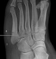
| 엑스레이에서 본 존스 골절 | |
| 전문 | 응급의료, 정형외과, 족부외과 |
| 증상 | 발 바깥쪽 중앙 부근의 통증, 타박상[2][3] |
| 통상적인 개시 | 서든[4] |
| 지속 | 치유까지[5] 6~12주 |
| 원인들 | 발끝이[6] 뾰족할 때 발을 안쪽으로 굽힌다. |
| 진단 방법 | 증상에 따라 엑스레이가[3] |
| 차동 진단 | 의사존 골절, 정상적인 성장판[3][7] |
| 치료 | 비중량 베어링, 깁스, 수술[5] |
존스 골절은 회복이 지연되거나 [4]결합이 되지 않는 것으로 알려진 저부와 중간부 사이의[8] 발의 다섯 번째 중족골의 특정 부위의 골절입니다.발 바깥쪽 [2]중앙 부근에 통증이 생깁니다.멍이 들거나 걷는 [3]데 어려움이 있을 수 있습니다.발병은 일반적으로 [4]갑작스럽다.
골절은 일반적으로 발가락이 뾰족하고 발이 [6][2]안쪽으로 구부러질 때 발생한다.이 움직임은 춤, 테니스 또는 [9][10]농구에서 발뒤꿈치가 그라운드에서 떨어져 있을 때 방향을 바꿀 때 발생할 수 있습니다.진단은 일반적으로 증상에 근거해 의심되며 엑스레이로 [3]확인된다.
초기 치료는 보통 최소 6주 [5]동안 걷지 않고 깁스를 합니다.이 기간이 지난 후에도 치유되지 않은 경우 추가로 6주간의 주조를 권장할 [5]수 있습니다.이 부위는 혈액공급이 잘 되지 않아 부러진 부분이 낫지 않아 수술이 필요할 [3]수 있다.운동선수나 뼈 조각이 분리되면 수술이 더 [5][8]빨리 고려될 수 있다.이 골절은 1902년 정형외과 의사 로버트 존스가 처음 설명했는데, 그는 춤을 [11][4]추다가 부상을 입었다.
진단.
존스 골절이 있는 사람은 골절이 발생했다는 것을 깨닫지 못할 수도 있다.진단에는 온전한 섬유상 브레비스 힘줄의 촉진과 제5중족골의 결절부에 원위하여 근위중족골의 축에 걸쳐 국소적인 압통을 나타내는 것이 포함된다.
진단 X선에는 전방 후방, 경사 및 측면 보기가 포함되며 발을 완전히 [citation needed]구부린 상태에서 촬영해야 합니다.
차동 진단


존스 골절만큼 문제가 되진 않지만 근위부 5번째 중족골절도 존재합니다.만약 골절이 중족간관절에 들어간다면 존스 골절입니다.그러나, 만약 그것이 슬관절 안으로 들어간다면, 그것은 섬유상뇌근의 인력으로 인한 박리골절일 가능성이 높다.제5중족골 밑부분의 박리골절은 때때로 "댄서 골절" 또는 "의사 존스 골절"이라고 불리며, 보통 수술하지 않는 [18]치료에 쉽게 반응한다.이 부위의 발달 "아포피시스"의 X선 모양은 골절과 유사할 수 있지만 골절은 아닙니다. 중족골의 2차 골화 중심입니다.이 [19]부위에서 청소년에게 나타나는 정상적인 소견입니다.해당 부위에 부상이 발생한 경우 의사는 종종 특정 방사선 단서를 해석하여 구별을 할 수 있습니다.이 위치의 박리 골절은 일반적으로 사용되지 않은 [19]아포피시스의 세로 방향과 비교하여 관절 외 및 가로 방향이다.
치료
캐스팅
초기 시술은 최소 [5]6주 동안 깁스를 하고 무게가 실리지 않습니다.이 기간이 지난 후에도 치유되지 않은 경우 추가로 6주 동안 주조하는 것이 좋습니다.[5]그러나 절반까지는 [2]캐스팅 후에도 낫지 않을 수 있습니다.
수술.
운동선수 또는 뼈 조각이 2mm 이상 떨어져 있는 경우 수술을 [5][8]고려할 수 있습니다.2009년부터 2015년까지 NFL 스카우팅 콤바인에 진출한 모든 선수를 대상으로 한 연구에서 존스 골절 발생률은 3.2%였으며 모두 금속 [20]나사로 골절을 수리하는 수술을 받았다.운동선수가 아닌 사람의 경우 깁스 [5]시술 후 치유되지 않는 한 수술이 권장되지 않을 수 있습니다.
예후
몇 가지 이유로, 존스 골절이 뭉치지 않을 수 있다.골절이 발생하는 간막골(Zone II)은 혈액 공급이 부족할 가능성이 있는 부위로, 두 혈액 공급 사이의 분수령 영역에 존재한다.이로 인해 치유가 저해될 수 있습니다.또한, 뼈에 붙어 있는 2개의 작은 근육과 섬유상완골, 섬유상완골 등 다양한 힘줄들이 있습니다.이것들은 골절을 떼어내고 [citation needed]치료를 방해할 수 있다.
구역 I과 III는 비교적 보장된 결합과 관련되어 있으며, 이 결합은 초기 고정화와 결합된 제한된 활동 제한만으로 이루어졌습니다.한편, 구역 II는 지연 또는 비결합과 관련되어 있으며, 따라서 [citation needed]내부 나사 고정과 같은 내부 고정화의 어떤 형태로든 이 부위의 골절을 고려해야 한다는 것이 일반적으로 합의되었다.
이러한 구역은 해부학적 및 X선 상에서 식별될 수 있으며, [21]이 분류의 임상적 유용성을 더합니다.외과적 개입은 그 자체로 완치를 보장하는 것은 아니며 합병증 발생률도 있다.문헌에 대한 다른 리뷰에서는 보수적이고 [22]비수술적인 치료가 비운동가들에게 허용 가능한 옵션이라는 결론을 내렸다.

다섯번째 중족골의 해부도.

3 존 설명
2 존 설명


레퍼런스
- ^ "5th Metatarsal". Emergency Care Institute, New South Wales. 2017-09-19.
- ^ a b c d Eltorai AE, Eberson CP, Daniels AH (2017). Orthopedic Surgery Clerkship: A Quick Reference Guide for Senior Medical Students. Springer. pp. 395–397. ISBN 9783319525679. Archived from the original on 2017-10-15.
- ^ a b c d e f "Toe and Forefoot Fractures". OrthoInfo - AAOS. June 2016. Archived from the original on 16 October 2017. Retrieved 15 October 2017.
- ^ a b c d Valderrabano V, Easley M (2017). Foot and Ankle Sports Orthopaedics. Springer. p. 430. ISBN 9783319157351. Archived from the original on 2017-10-15.
- ^ a b c d e f g h i Bica D, Sprouse RA, Armen J (February 2016). "Diagnosis and Management of Common Foot Fractures". American Family Physician. 93 (3): 183–91. PMID 26926612.
- ^ a b Dähnert W (2011). Radiology Review Manual. Lippincott Williams & Wilkins. p. 96. ISBN 9781609139438. Archived from the original on 2017-10-15.
- ^ Conaghan PG, O'Connor P, Isenberg DA (2010). Musculoskeletal Imaging. OUP Oxford. p. 231. ISBN 9780191575273. Archived from the original on 2017-10-15.
- ^ a b c Joel A. DeLisa; Bruce M. Gans; Nicholas E. Walsh (2005). Physical Medicine and Rehabilitation: Principles and Practice. Lippincott Williams & Wilkins. pp. 881–. ISBN 978-0-7817-4130-9. Archived from the original on 2017-01-07.
- ^ Mattu A, Chanmugam AS, Swadron SP, Tibbles C, Woolridge D, Marcucci L (2012). Avoiding Common Errors in the Emergency Department. Lippincott Williams & Wilkins. p. 790. ISBN 9781451152852. Archived from the original on 2017-10-16.
- ^ Lee E (2017). Pediatric Radiology: Practical Imaging Evaluation of Infants and Children. Lippincott Williams & Wilkins. p. Chapter 24. ISBN 9781496380272. Archived from the original on 2017-10-15.
- ^ Jones R (June 1902). "I. Fracture of the Base of the Fifth Metatarsal Bone by Indirect Violence". Annals of Surgery. 35 (6): 697–700.2. PMC 1425723. PMID 17861128.
- ^ Bica D, Sprouse RA, Armen J (February 2016). "Diagnosis and Management of Common Foot Fractures". American Family Physician. 93 (3): 183–91. PMID 26926612.
- ^ "5th Metatarsal". Emergency Care Institute, New South Wales. 2017-09-19.
- ^ "Toe and Forefoot Fractures". OrthoInfo - AAOS. June 2016. Archived from the original on 16 October 2017. Retrieved 15 October 2017.
- ^ a b Silbergleit R. "Foot Fracture". Medscape.com. Retrieved 19 October 2011.
- ^ Deniz G, Kose O, Guneri B, Duygun F (May 2014). "Traction apophysitis of the fifth metatarsal base in a child: Iselin's disease". BMJ Case Reports. 2014 (may14 4): bcr2014204687–bcr2014204687. doi:10.1136/bcr-2014-204687. PMID 24832713.
- ^ Nwawka OK, Hayashi D, Diaz LE, Goud AR, Arndt WF, Roemer FW, et al. (October 2013). "Sesamoids and accessory ossicles of the foot: anatomical variability and related pathology". Insights Into Imaging. 4 (5): 581–93. doi:10.1007/s13244-013-0277-1. PMC 3781258. PMID 24006205.
- ^ "Toe and Forefoot Fractures/Fifth Metatarsal Fractures". orthoinfo.aaos.org. American Academy of Orthopedic Surgeons. Retrieved November 3, 2021.
- ^ a b Saber, Mohamed; Sharma, Rohit (March 26, 2021). "Apophysis of the proximal 5th metatarsal". radiopaedia.org. Radiopedia.org. Retrieved November 3, 2021.
- ^ Spang, Robert C. (August 2018). "Jones Fractures Identified at the National Football League Scouting Combine: Assessment of Prognostic Factors, Computed Tomography Findings, and Initial Career Performance". Orthopaedic Journal of Sports Medicine. 6 (8). doi:10.1177/2325967118790740. ISSN 2325-9671. PMC 6113739. PMID 30182027.
- ^ Polzer H, Polzer S, Mutschler W, Prall WC (October 2012). "Acute fractures to the proximal fifth metatarsal bone: development of classification and treatment recommendations based on the current evidence". Injury. 43 (10): 1626–32. doi:10.1016/j.injury.2012.03.010. PMID 22465516.
- ^ Dean BJ, Kothari A, Uppal H, Kankate R (August 2012). "The jones fracture classification, management, outcome, and complications: a systematic review". Foot & Ankle Specialist. 5 (4): 256–9. doi:10.1177/1938640012444730. PMID 22547534. S2CID 37169110.


