피부내시경
Dermatoscopy| 피부내시경 | |
|---|---|
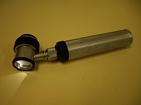
 몰딩 오일 피부경. | |
| 전문 | 피부과 |
| 메슈 | D046169 |
피부진단은 피부과로 피부병변을 검사하는 것이다.
진경 검사 또는 발광 현미경 검사로도 알려져 있으며, 피부 표면 반사에 의해 방해받지 않는 피부 병변을 검사할 수 있다.피부경은 돋보기와 광원(극화 또는 비극화), 투명판, 때로는 기기와 피부 사이에 액체 매질로 구성되어 있다.영상이나 비디오 클립을 디지털로 캡처하거나 처리할 때, 기기를 디지털 후광 피부시술이라고 할 수 있다.
이 기술은 피부과 의사나 피부암 전문의가 양성과 악성(암) 병변을 구분하는데 유용하며, 특히 흑색종 진단에 유용하다.
피부진단의 종류
피부경은 전진 광원과 돋보기(보통 10배경(보통 10배 확대)심내시경에는 다음과 같은 세 가지 주요 모드가 있다.[1]
편광은 더 깊은 피부 구조를 시각화할 수 있는 반면, 비편광은 피상적인 피부에 대한 정보를 제공한다.대부분의 현대적인 피부과에서는 사용자가 두 가지 모드 사이를 전환할 수 있게 하는데, 이것은 상호 보완적인 정보를 제공한다.
피부진단의 장점
피부내시경 전문가인 의사들이 있어 전문 교육을 받지 않은 의사들보다 흑색종에 대한 진단 정확도가 월등히 우수하다.[2]따라서 육안검사에 비해 특이성(양성으로 올바르게 진단된 비멜라노마스의 비율)뿐만 아니라 민감도(멜라노마스의 검출)도 상당히 개선되었다.피부진단에 의한 정확도는 육안검사에 비해 민감도의 경우 최대 20%까지, 특수성의 경우 최대 10%까지 높였다.[3][4]따라서 피부진단을 사용하면 특이성이 증가하여 양성 병변의 불필요한 수술 절개 빈도가 감소한다.[5][6]
피부검진 적용
- 피부검사의 대표적인 적용은 흑색종 조기 발견(위 참조)이다.
- 흑색종이 의심스러운 피부병변을 모니터링하는 데 디지털 피부진찰(비디오메타내시경)이 사용된다.디지털 피부과 진단 영상이 저장되며 환자의 다음 방문 때 얻은 영상과 비교된다.그러한 병변의 의심스러운 변화는 절제를 위한 지표다.시간이 지남에 따라 변하지 않는 피부 병변은 양성인 것으로 간주된다.[7][8]디지털 더모시경을 위한 일반적인 시스템은 Fotofinder, Molemax, DermoGenius, Easyscan 또는 하이네이다.
- 피부 종양의 진단에 도움을 주는 것 - 기저 세포암,[9] 편평한 세포암,[10] 원통형 세포암,[11] 피부색소, 혈관종, 지루성 각질증 및 기타 많은 일반적인 피부 종양들은 고전적인 피부경 검사 결과를 가지고 있다.[12]
- 딱지 및 치골의 진단에 도움이 된다.인디아 잉크로 피부를 물들임으로써 피부시경은 굴 속의 진드기 위치를 식별하는 데 도움을 줄 수 있으며, 이로 인해 혈당 굴의 스크래핑이 용이해진다.음핵을 확대함으로써, 그것은 작은 곤충들을 보기 어려운 사람들을 신속하게 진단할 수 있다.[13][14]
- 사마귀 진단에 도움이 된다.의사가 사마귀의 구조를 시각화할 수 있게 함으로써, 그것을 옥수수, 집, 외상, 또는 이물질과 구별할 수 있게 한다.치료 후기 사마귀 검사를 통해 사마귀 구조를 시각화하기가 어려워 치료가 조기에 중단되지 않도록 한다.
- 진균 감염의 진단에 도움이 된다."검은 점" 티네아, 또는 티네아 캡티염(엉덩이 두피 감염)을 탈모증 아레아타와 구별하기 위해서입니다.[15]
- 탈모증후군,[16] 여성 안드로겐성 탈모증,[17] 모닐트릭스,[18] 네더톤 증후군,[19] 양털모발증후군 등 모발과 두피질환 진단에 도움이 된다.[20]모발과 두피의 심내시경 검사를 삼차경이라고 한다.[21][22]
- 피부암을 정의하기 어려운 수술 여백의 결정.예를 들면 보웬병, 피상적인 기저세포암, 렌티고 말라이다.이 종양들은 여백이 매우 불분명하다.외과의사가 종양의 실제 범위를 정확하게 파악할 수 있게 함으로써, 반복 수술은 종종 감소한다.
- 악성 흑색종 또는 접합성 흑색종과 티네아 니그라의 구별.[23]
역사
피부 표면 현미경 검사는 1663년 콜하우스에 의해 시작되었고, 1878년 에른스트 아베에 의해 몰입 오일이 첨가되면서 개선되었다.독일의 피부과 전문의 요한 사피에가 기구에 내장된 광원을 추가했다.골드만은 피부과 의사로는 최초로 '더마스카피'라는 용어를 쓰고 피부과로 색소침착 피부병변을 평가한 바 있다.
1989년 루드비히스-맥시밀리안-뮌헨 대학의 피부과 의사들이 더모시경을 위한 새로운 장치를 개발했다.오토 브라운-팔코 교수팀이 의료기기 제조업체 하이네 옵토테크닉과 손잡고 피부과 시술을 개발했는데, 이 시계는 손으로 들고 할로겐 램프로 조명이 켜졌다.10배 확대된 무채색 렌즈도 선보였다.빛 반사를 줄이기 위해 병변은 침전 오일로 덮여 있었다.이 피부시경은 색소성 피부병변을 더 빠르고 쉽게 진단하는데 도움을 주었다.이 접근방식은 뮌헨 대학의 피부과 알레르기와학과의 빌헬름 스톨츠 외 연구진에 의해 확인되었고 "랜싯"(1989)에 발표되었다.[24]
비엔나 의과대학에서는 교차 폴라화 기반의 피부과가 발명되어 특허를 받았으며, MoleMax™-장치와 같은 디지털 피부과나 FotoFinder에 의해 추가로 사용되는 방법론이 개발되었다.2001년에 이어, 캘리포니아의 의료기기 제조업체인 3Gen이 최초로 극성을 띠는 핸드헬드 피부경인 DermLite를 선보였다.편광 조명은 교차 폴라화 뷰어와 결합되어 피부 표면 반사를 감소(폴라화)하므로, 몰입 액체를 사용하지 않고도 피부 구조(탈극화 빛)를 시각화할 수 있다.따라서 의사가 각 병변을 검사하기 전에 더 이상 침전 오일, 알코올 또는 물을 피부에 바르지 않아도 되기 때문에 여러 병변을 검사하는 것이 더 편리하다.편광피부과 마케팅으로 전 세계 의사들 사이에서 피부진단이 인기를 끌었다.편광광 피부과에서 생산되는 영상은 기존의 피부 접촉 유리 피부과에서 생산되는 영상과 약간 다르지만 유리 접촉판에 의한 피부 압축을 통해 혈관 패턴을 잠재적으로 놓치지 않는 등 확실한 장점이 있다.[25]
상당히 표준화된 이미지와 임상 피부과에 비해 진단량이 제한되어 있어 피부과 영상은 자동화된 의료 영상 분석의 관심의 중심이 되었다.과거 수십 년 동안 컴퓨터 비전 알고리즘과 하드웨어 기반 방법이 사용된 반면, HAM10000과[28] 같은 대규모 표준화된 공공 이미지 컬렉션은 경련 신경 네트워크의 적용을 가능하게 했다.[27]후자의 접근방식은 이제 더 큰/국제적인,[30][31][32] 그리고 더 작은/지역적인 실험에서 인간 수준의 정확성에 대한 실험 증거를 보여주었지만,[34][35] 이 적용은 논쟁이 없는 것은 아니다.[37]
참조
- ^ Argenziano, G; Soyer, HP (July 2001). "Dermoscopy of pigmented skin lesions--a valuable tool for early diagnosis of melanoma". The Lancet. Oncology. 2 (7): 443–9. doi:10.1016/s1470-2045(00)00422-8. PMID 11905739.
- ^ Lorentzen, H; Weismann, K; Petersen, CS; Larsen, FG; Secher, L; Skødt, V (1999). "Clinical and dermatoscopic diagnosis of malignant melanoma. Assessed by expert and non-expert groups". Acta Dermato-venereologica. 79 (4): 301–4. doi:10.1080/000155599750010715. PMID 10429989.
- ^ Vestergaard, ME; Macaskill, P; Holt, PE; Menzies, SW (2008). "Dermoscopy compared with naked eye examination for the diagnosis of primary melanoma: a meta-analysis of studies performed in a clinical setting". British Journal of Dermatology. 159 (3): 669–76. doi:10.1111/j.1365-2133.2008.08713.x. PMID 18616769. S2CID 46404467.Argenziano, G; Fabbrocini, G; Carli, P; De Giorgi, V; Sammarco, E; Delfino, M (1998). "Epiluminescence microscopy for the diagnosis of doubtful melanocytic skin lesions. Comparison of the ABCD rule of dermatoscopy and a new 7-point checklist based on pattern analysis". Archives of Dermatology. 134 (12): 1563–70. doi:10.1001/archderm.134.12.1563. PMID 9875194.
- ^ Ascierto, P.A.; Palmieri, G.; Celentano, E.; Parasole, R.; Caraco, C.; Daponte, A.; Chiofalo, M.G.; Melucci, M.T.; Mozzillo, N.; Satriano, R.A.; Castello, G. (2000). "Sensitivity and specificity of epiluminescence microscopy: evaluation on a sample of 2731 excised cutaneous pigmented lesions". British Journal of Dermatology. 142 (5): 893–8. doi:10.1046/j.1365-2133.2000.03468.x. PMID 10809845. S2CID 70893256.http://www.bcbstx.com/provider/pdf/medicalpolicies/medicine/201-023.pdf[출처?][영구적 데드링크]
- ^ Bono, A; Bartoli, C; Cascinelli, N; Lualdi, M; Maurichi, A; Moglia, D; Tragni, G; Tomatis, S; Marchesini, R (2002). "Melanoma detection. A prospective study comparing diagnosis with the naked eye, dermatoscopy and telespectrophotometry". Dermatology. 205 (4): 362–6. doi:10.1159/000066436. PMID 12444332. S2CID 25348202.
- ^ "Crutchfield Dermatology". Crutchfield Dermatology. Archived from the original on 2016-10-11. Retrieved 2010-05-12.
- ^ Argenziano, G; Mordente, I; Ferrara, G; Sgambato, A; Annese, P; Zalaudek, I (2008). "Dermoscopic monitoring of melanocytic skin lesions: clinical outcome and patient compliance vary according to follow-up protocols". The British Journal of Dermatology. 159 (2): 331–6. doi:10.1111/j.1365-2133.2008.08649.x. PMID 18510663. S2CID 32027237.
- ^ Roma, Paolo; Savarese, Imma; Martino, Antonia; Martino, Domenico; Annese, Pietro; Capoluongo, Patrizio; Mordente, Ines; Nicolino, Rachele; Zalaudek, Iris; Argenziano, Giuseppe (2007). "Slow-growing melanoma: Report of five cases". Journal of Dermatological Case Reports. 1 (1): 1–3. doi:10.3315/jdcr.2007.1.1001. PMC 3157767. PMID 21886697.
- ^ Scalvenzi, M; Lembo, S; Francia, MG; Balato, A (2008). "Dermoscopic patterns of superficial basal cell carcinoma". International Journal of Dermatology. 47 (10): 1015–8. doi:10.1111/j.1365-4632.2008.03731.x. PMID 18986346. S2CID 205394964.
- ^ Felder, S; Rabinovitz, H; Oliviero, M; Kopf, A (2006). "Dermoscopic differentiation of a superficial basal cell carcinoma and squamous cell carcinoma in situ". Dermatologic Surgery. 32 (3): 423–5. doi:10.1111/j.1524-4725.2006.32085.x. PMID 16640692.
- ^ Sicinska, Justyna; Rakowska, Adriana; Czuwara-Ladykowska, Joanna; Mroz, Andrzej; Lipinski, Marcin; Nasierowska-Guttmejer, Anna; Sikorska, Jolanta; Sklinda, Katarzyna; Slowinska, Monika; Kowalska-Oledzka, Elzbieta; Walecka, Irena; Walecki, Jerzy; Rudnicka, Lidia (2007). "Cylindroma transforming into basal cell carcinoma in a patient with Brooke-Spiegler syndrome". Journal of Dermatological Case Reports. 1 (1): 4–9. doi:10.3315/jdcr.2007.1.1002. PMC 3157764. PMID 21886698.
- ^ Campos-Do-Carmo, G; Ramos-E-Silva, M (2008). "Dermoscopy: basic concepts". International Journal of Dermatology. 47 (7): 712–9. doi:10.1111/j.1365-4632.2008.03556.x. PMID 18613881. S2CID 205394507.
- ^ Wu, Ming-Yun; Hu, Shu-Lin; Hsu, Che-Hao (June 2008). "Use of Non-contact Dermatoscopy in the Diagnosis of Scabies" (PDF). Dermatol Sinica: 112–4. Archived from the original (PDF) on 2011-07-09. Retrieved 2009-09-28.
- ^ Chuh, A; Lee, A; Wong, W; Ooi, C; Zawar, V (2007). "Diagnosis of Pediculosis pubis: a novel application of digital epiluminescence dermatoscopy". Journal of the European Academy of Dermatology and Venereology. 21 (6): 837–8. doi:10.1111/j.1468-3083.2006.02040.x. PMID 17567326. S2CID 34621172.
- ^ Slowinska, M; Rudnicka, L; Schwartz, RA; Kowalska-Oledzka, E; Rakowska, A; Sicinska, J; Lukomska, M; Olszewska, M; Szymanska, E (2008). "Comma hairs: a dermatoscopic marker for tinea capitis: a rapid diagnostic method". Journal of the American Academy of Dermatology. 59 (5 Suppl): S77–9. doi:10.1016/j.jaad.2008.07.009. PMID 19119131.
- ^ Inui, S; Nakajima, T; Itami, S (2008). "Significance of dermoscopy in acute diffuse and total alopecia of the female scalp: review of twenty cases". Dermatology. 217 (4): 333–6. doi:10.1159/000155644. PMID 18799878. S2CID 19963257.
- ^ Rakowska, A.; et al. (2008). "Trichoscopy criteria for diagnosing female androgenic alopecia". Nature Precedings. doi:10.1038/npre.2008.1913.1. hdl:10101/npre.2008.1913.1.
- ^ Rakowska, A; Slowinska, M; Czuwara, J; Olszewska, M; Rudnicka, L (2007). "Dermoscopy as a tool for rapid diagnosis of monilethrix". Journal of Drugs in Dermatology. 6 (2): 222–4. PMID 17373184.
- ^ Rakowska, A; Kowalska-Oledzka, E; Slowinska, M; Rosinska, D; Rudnicka, L (2009). "Hair shaft videodermoscopy in netherton syndrome". Pediatric Dermatology. 26 (3): 320–2. doi:10.1111/j.1525-1470.2008.00778.x. PMID 19706096. S2CID 42660583.
- ^ Rakowska, Adriana; Slowinska, Monika; Kowalska-Oledzka, Elzbieta; Rudnicka, Lidia (2008). "Trichoscopy in genetic hair shaft abnormalities". Journal of Dermatological Case Reports. 2 (2): 14–20. doi:10.3315/jdcr.2008.1009. PMC 3157768. PMID 21886705.
- ^ Rudnicka L, Olszewska M, Rakowska A, Kowalska-Oledzka E, Slowinska M (July 2008). "Trichoscopy: a new method for diagnosing hair loss". Journal of Drugs in Dermatology. 7 (7): 651–4. PMID 18664157.
- ^ Rakowska A, Slowinska M, Kowalska-Oledzka E, Rudnicka L (2008). "Trichoscopy (hair and scalp videodermoscopy) in the healthy female. Method standardization and norms for measurable parameters". J Dermatol Case Rep. 3 (1): 14–19. doi:10.3315/jdcr.2008.1021. PMC 3157785. PMID 21886722.
- ^ Guarenti, IM; Almeida HL, Jr; Leitão, AH; Rocha, NM; Silva, RM (2013). "Scanning electron microscopy of tinea nigra". Anais Brasileiros de Dermatologia. 89 (2): 334–6. doi:10.1590/abd1806-4841.20142780. PMC 4008070. PMID 24770516.
- ^ Stolz, Wilhelm (1989). "Skin Surface Microscopy". The Lancet. 334 (8667): 864–865. doi:10.1016/s0140-6736(89)93027-4. PMID 2571785. S2CID 5459081.
- ^ "Non-contact dermatoscopic device with full polarization control and liquid lens based autofocus function" (PDF). www.dgao-proceedings.de. Retrieved 2019-01-16.
- ^ Dreiseitl, S; Binder, M; Hable, K; Kittler, H (June 2009). "Computer versus human diagnosis of melanoma: evaluation of the feasibility of an automated diagnostic system in a prospective clinical trial". Melanoma Research. 19 (3): 180–4. doi:10.1097/CMR.0b013e32832a1e41. PMID 19369900. S2CID 3095768.
- ^ Dick, V; Sinz, C; Mittlböck, M; Kittler, H; Tschandl, P (19 June 2019). "Accuracy of Computer-Aided Diagnosis of Melanoma: A Meta-analysis". JAMA Dermatology. 155 (11): 1291–1299. doi:10.1001/jamadermatol.2019.1375. PMC 6584889. PMID 31215969.
- ^ Tschandl, P; Rosendahl, C; Kittler, H (14 August 2018). "The HAM10000 dataset, a large collection of multi-source dermatoscopic images of common pigmented skin lesions". Scientific Data. 5: 180161. arXiv:1803.10417. Bibcode:2018NatSD...580161T. doi:10.1038/sdata.2018.161. PMC 6091241. PMID 30106392.
- ^ Tschandl, P; Codella, N; Akay, BN; Argenziano, G; Braun, RP; Cabo, H; Gutman, D; Halpern, A; Helba, B; Hofmann-Wellenhof, R; Lallas, A; Lapins, J; Longo, C; Malvehy, J; Marchetti, MA; Marghoob, A; Menzies, S; Oakley, A; Paoli, J; Puig, S; Rinner, C; Rosendahl, C; Scope, A; Sinz, C; Soyer, HP; Thomas, L; Zalaudek, I; Kittler, H (July 2019). "Comparison of the accuracy of human readers versus machine-learning algorithms for pigmented skin lesion classification: an open, web-based, international, diagnostic study". The Lancet. Oncology. 20 (7): 938–947. doi:10.1016/S1470-2045(19)30333-X. PMC 8237239. PMID 31201137.
- ^ Tschandl, P; Rosendahl, C; Akay, BN; Argenziano, G; Blum, A; Braun, RP; Cabo, H; Gourhant, JY; Kreusch, J; Lallas, A; Lapins, J; Marghoob, A; Menzies, S; Neuber, NM; Paoli, J; Rabinovitz, HS; Rinner, C; Scope, A; Soyer, HP; Sinz, C; Thomas, L; Zalaudek, I; Kittler, H (1 January 2019). "Expert-Level Diagnosis of Nonpigmented Skin Cancer by Combined Convolutional Neural Networks". JAMA Dermatology. 155 (1): 58–65. doi:10.1001/jamadermatol.2018.4378. PMC 6439580. PMID 30484822.
- ^ Marchetti, MA; Codella, NCF; Dusza, SW; Gutman, DA; Helba, B; Kalloo, A; Mishra, N; Carrera, C; Celebi, ME; DeFazio, JL; Jaimes, N; Marghoob, AA; Quigley, E; Scope, A; Yélamos, O; Halpern, AC; International Skin Imaging, Collaboration. (February 2018). "Results of the 2016 International Skin Imaging Collaboration International Symposium on Biomedical Imaging challenge: Comparison of the accuracy of computer algorithms to dermatologists for the diagnosis of melanoma from dermoscopic images". Journal of the American Academy of Dermatology. 78 (2): 270–277.e1. doi:10.1016/j.jaad.2017.08.016. PMC 5768444. PMID 28969863.
- ^ Haenssle, HA; Fink, C; Toberer, F; Winkler, J; Stolz, W; Deinlein, T; Hofmann-Wellenhof, R; Lallas, A; Emmert, S; Buhl, T; Zutt, M; Blum, A; Abassi, MS; Thomas, L; Tromme, I; Tschandl, P; Enk, A; Rosenberger, A; Reader Study Level I and Level II, Groups. (January 2020). "Man against machine reloaded: performance of a market-approved convolutional neural network in classifying a broad spectrum of skin lesions in comparison with 96 dermatologists working under less artificial conditions". Annals of Oncology. 31 (1): 137–143. doi:10.1016/j.annonc.2019.10.013. PMID 31912788.
- ^ Tschandl, P; Kittler, H; Argenziano, G (September 2017). "A pretrained neural network shows similar diagnostic accuracy to medical students in categorizing dermatoscopic images after comparable training conditions". The British Journal of Dermatology. 177 (3): 867–869. doi:10.1111/bjd.15695. PMID 28569993. S2CID 207076965.
- ^ Yu, C; Yang, S; Kim, W; Jung, J; Chung, KY; Lee, SW; Oh, B (2018). "Acral melanoma detection using a convolutional neural network for dermoscopy images". PLOS ONE. 13 (3): e0193321. Bibcode:2018PLoSO..1393321Y. doi:10.1371/journal.pone.0193321. PMC 5841780. PMID 29513718.
- ^ Esteva, A; Kuprel, B; Novoa, RA; Ko, J; Swetter, SM; Blau, HM; Thrun, S (2 February 2017). "Dermatologist-level classification of skin cancer with deep neural networks". Nature. 542 (7639): 115–118. Bibcode:2017Natur.542..115E. doi:10.1038/nature21056. PMC 8382232. PMID 28117445.
- ^ Lallas, A; Argenziano, G (October 2018). "Artificial intelligence and melanoma diagnosis: ignoring human nature may lead to false predictions". Dermatology Practical & Conceptual. 8 (4): 249–251. doi:10.5826/dpc.0804a01. PMC 6246056. PMID 30479851.
- ^ Navarrete-Dechent, C; Dusza, SW; Liopyris, K; Marghoob, AA; Halpern, AC; Marchetti, MA (October 2018). "Automated Dermatological Diagnosis: Hype or Reality?". The Journal of Investigative Dermatology. 138 (10): 2277–2279. doi:10.1016/j.jid.2018.04.040. PMC 7701995. PMID 29864435.


