PSMD2
PSMD226S 프로테아솜 비 ATPase 규제 서브 유닛 2(시스템 명명법 26S Proteasome Regulation Subunit Rpn1)는 인간에서 PSMD2 유전자에 의해 암호화된 효소다.[5][6]
구조
유전자 발현상
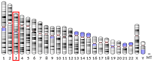
PSMD2 유전자는 기질 인식과 결합을 담당하는 19S 조절기 베이스의 비 ATPase 하위 유닛을 인코딩한다.[6]PSMD2 유전자는 19S 레귤레이터 리드의 비 ATPase 하위 결합 중 하나를 암호화한다.이 서브유닛은 프로테아솜 기능 참여 외에도 종양 괴사 인자 타입 1 수용체와 상호작용하기 때문에 TNF 신호전달 경로에도 참여할 수 있다.1번 염색체에서 유사 유전자가 확인되었다.[6]인간 PSMD2 유전자는 23개의 exon을 가지고 있으며 3q27.1 염색체 밴드에 위치한다.인간 단백질 26S 프로테아솜 비ATPase 규제 소단위 2는 크기가 100kDa이며 909개의 아미노산으로 구성된다.이 단백질의 계산된 이론적 pI는 5.10이다.대체 스플라이싱에 의해 두 가지 표현식 이소폼이 생성되는데, 이 과정에서 아미노산 시퀀스의 1-130 또는 1-163이 누락된다.
복합 조립체
26S 프로테아솜 콤플렉스는 보통 20S 코어 입자(CP 또는 20S 프로테아솜)와 배럴 모양의 20S 한쪽 또는 양쪽에 1개 또는 2개의 19S 규제 입자(RP 또는 19S 프로테아솜)로 구성된다.CP와 RP는 서로 다른 구조적 특성과 생물학적 기능을 가지고 있다.간단히 말해서, 20S 하위 단지는 카스파제 유사, 트립신 유사, 키모트립신 유사 활동을 포함한 세 가지 유형의 단백질 분해 활동을 제시한다.20S 서브유닛의 4개의 스택 링에 의해 형성된 챔버의 내측에 위치한 이러한 단백질 분해 활성 사이트는 무작위 단백질-엔자임 조우 및 제어되지 않은 단백질 저하를 방지한다.19S 규제 입자는 유비퀴틴 라벨이 부착된 단백질을 분해 기질로 인식하고, 단백질을 선형까지 펼치며, 20S 코어 입자의 문을 열고, 변전체를 프로톨리틱 챔버로 안내할 수 있다.그러한 기능적 복잡성을 충족시키기 위해 19S 규제 입자는 최소 18개의 구성 하위 단위를 포함한다.이러한 서브유닛은 서브유닛의 ATP 의존성, ATP 종속 서브유닛 및 ATP 독립 서브유닛에 근거한 두 가지 등급으로 분류할 수 있다.이 다단위 단지의 단백질 상호작용과 위상학적 특성에 따라 19S 규제 입자는 베이스와 리드 서브콤플렉스로 구성된다.베이스는 6개의 AAA ATPASes(Subunit Rpt1-6, 체계적 명명법)와 4개의 비 ATPase 서브유닛(Rpn1, Rpn2, Rpn10, Rpn13)으로 구성된 링으로 구성된다.따라서 단백질 26S 프로테아솜 비 ATPase 규제 서브 유닛 2(Rpn1)는 19S 규제 입자의 기본 하위 콤플렉스를 형성하는 데 필수적인 구성요소다.전통적으로, Rpn1과 Rpn2는 기지 서브 콤플렉스 중심에 거주하고 6개의 AAA ATPAS(Rpt 1-6)로 둘러싸인 것으로 간주되었다.그러나, 최근의 조사는 극저온 현미경, X선 결정학, 잔류물 특정 화학 교차 링크 및 몇 가지 단백질학 기법의 데이터를 결합한 통합 접근방식을 통해 19S 기반 대체 구조를 제공한다.Rpn2는 ATPase 링의 측면에 위치한 강체 단백질로 뚜껑과 베이스 사이의 연결부로 지지된다.Rpn1은 순응적으로 가변적이며, ATPase 링의 주변부에 위치한다.유비퀴틴 수용체 Rpn10과 Rpn13은 19S 콤플렉스 원위부 더 멀리 위치해 있어 조립 과정에서 늦게 콤플렉스에 포섭된 것을 알 수 있다.[7]
함수
세포내 단백질 분해의 약 70%를 담당하는 분해기계로서 프로테아솜 콤플렉스(26S 프로테아솜)는 세포 프로테오메의 동태성을 유지하는 데 중요한 역할을 한다.[8]따라서 잘못 접힌 단백질과 손상된 단백질은 새로운 합성을 위해 아미노산을 재활용하기 위해 지속적으로 제거되어야 한다; 동시에, 일부 핵심 규제 단백질은 선택적 저하를 통해 생물학적 기능을 수행하며, 더욱이 단백질은 MHC 등급 I 항원 발현을 위해 펩타이드로 소화된다.공간적 및 시간적 단백질분해를 통해 생물학적 과정에서 이처럼 복잡한 요구를 충족시키기 위해서는 단백질 기판을 잘 통제된 방식으로 인식하고, 모집하고, 결국에는 가수분해해야 한다.따라서 19S 규제 입자는 이러한 기능적 과제를 해결하기 위한 일련의 중요한 기능을 포함한다.단백질을 지정된 기질로 인식하기 위해 19S 콤플렉스는 특수 분해 태그인 유비쿼터스비닐화(ubiquititial tag)로 단백질을 인식할 수 있는 서브유닛을 갖췄다.또한 19S와 20S 입자의 연계를 용이하게 하기 위해 뉴클레오티드(예: ATP)와 결합할 수 있는 서브유닛(subunit)을 가지고 있으며, 20S 단지의 하부 정문을 형성하는 알파 서브유닛 C-단자의 확인변경을 유발한다.Rpn1은 19S 규제 입자의 필수적인 하위 단위 중 하나이며, 그것은 "베이스" 하위 복합체의 핵심을 형성한다.Rpn10과의 연관성은 제3 서브 유닛인 Rpn2에 의해 안정화되지만, 다른 19S 서브 유닛 Rpn10의 도킹 위치를 중앙 솔레노이드 부분에 제공한다.[9]Rpn2는 19S 복합 조립에서 중요한 역할 외에도 유비퀴티비닐화 기질 밀거래 셔틀에 도킹 위치를 제공한다.대부분의 셔틀은 UBL(Universitin 유사 도메인)을 통해 프로테아솜에 부착되며, C-단자 폴리우비퀴틴 결합 도메인에서 기판 화물을 하역한다.글릭먼 등의 최근 조사에 따르면 Rad23과 Dsk2라는 두 개의 셔틀 단백질이 서브유닛 Rpn1에 내장된 두 개의 서로 다른 수용체 부위에서 도킹하는 것으로 밝혀졌다.[9]
임상적 유의성
프로테아솜과 그 서브유닛은 적어도 두 가지 이유로 임상적으로 중요한데, (1) 손상된 복합체 결합 또는 기능장애 프로테아솜은 특정 질병의 근본적인 병태생리와 관련될 수 있으며, (2) 치료적 개입의 약물 대상으로 악용될 수 있다.보다 최근에는 새로운 진단 마커와 전략의 개발을 위한 프로테아솜을 고려하는 노력이 더 많이 이루어지고 있다.프로테아좀의 병태생리학에 대한 개선되고 포괄적인 이해는 향후 임상적 응용으로 이어져야 한다.
프로테아솜은 유비퀴틴-단백질 시스템(UPS)과 해당 세포 단백질 품질 관리(PQC)의 중추적 구성 요소를 형성한다.단백질 편재와 그에 따른 프로테아솜에 의한 단백질 분해와 분해는 세포 주기, 세포 성장과 분화, 유전자 전사, 신호 전달 및 세포 사멸의 조절에 있어 중요한 메커니즘이다.[11]그 후 단백질 복합체 조립 및 기능이 손상되면 단백질 분해 활동이 감소하고 단백질 종들이 손상되거나 잘못 접히는 현상이 발생한다.이러한 단백질이 축적되면 신경퇴행성질환,[12][13] 심혈관질환,[14][15][16] 염증반응 및 자가면역질환,[17] 전신 DNA손상반응에서 병생성과 표현특성에 기여하여 악성종양으로 이어질 수 있다.[18]
몇몇 임상 실험 연구 성격의 UPS수차와 규제 완화 등 몇몇이고myodegenerative 신경 퇴행성 질환의 발병에 기여하고의 disease,[21]근 위축성 측색 경화(근 위축성 측색 경화증)[21]헌팅턴 disease,[20]Creutz다 알츠하이머 disease,[19]파킨슨 병 disease[20]등을 표시했습니다.feldt–Jakob disease,[22]그리고 운동 뉴런 질환, 폴리글루타민(PolyQ) 질환, 근위축성[23] 및 치매와 관련된 몇 가지 희귀한 형태의 신경퇴행성 질환이 있다.[24]유비퀴틴-단백질계통(UPS)의 일부로서 프로테아솜은 심장 단백질 동점증을 유지하여 심장 허혈성 부상,[25] 심실 비대증[26], 심부전 등에 큰 역할을 한다.[27]게다가, UPS가 악성 변형에 필수적인 역할을 한다는 증거가 축적되고 있다.UPS 단백질 분해는 암의 발병에 중요한 자극 신호에 대한 암세포의 반응에 중요한 역할을 한다.따라서 p53, c-준, c-Fos, NF-bB, c-Myc, HIF-1α, MATα2, STAT3, 스테롤 조절 요소 결합 단백질 및 안드로겐 수용체와 같은 전사 인자의 저하로 인한 유전자 발현이 모두 UPS에 의해 제어되어 다양한 악성종양 개발에 관여한다.[28]Moreover, the UPS regulates the degradation of tumor suppressor gene products such as adenomatous polyposis coli (APC) in colorectal cancer, retinoblastoma (Rb). and von Hippel–Lindau tumor suppressor (VHL), as well as a number of proto-oncogenes (Raf, Myc, Myb, Rel, Src, Mos, ABL).UPS도 염증반응 규제에 관여하고 있다.이러한 활동은 대개 TNF-α, IL-β, IL-8, 접착분자(ICAM-1, VCAM-1, P-selectin)와 프로스타글란딘과 질산화물(NO)과 같은 프로테아그란딘의 발현을 더욱 조절하는 NF-164B의 활성화에 있어서 프로테아솜의 역할에 기인한다.[17]또한 UPS는 주로 사이클린의 단백질 분해와 CDK 억제제의 저하를 통해 백혈구 증식의 조절자로서 염증 반응에도 역할을 한다.[29]마지막으로, SLE, Sögren 증후군, 류마티스 관절염(RA)을 앓고 있는 자가면역질환자들은 임상 바이오마커로 응용할 수 있는 순환 프로테아솜을 주로 나타낸다.[30]
PSMD2에 의해 암호화된 26S 단백질이 아닌 ATPase 규제 소단위 2(Rpn1)는 전이적 표현형 및 폐암에서의 좋지 않은 예후 획득과 관련된 서명의 중요한 구성 요소로 확인되었다.[31]PSMD2의 녹다운은 프로테아솜 활동을 감소시키고, 폐암 세포 라인의 성장 억제와 사멸을 유도하는 것으로 밝혀졌다.siRNA 매개 PSMD2 억제의 이러한 영향은 p21의 유도뿐만 아니라 인광 AKT와 p38 사이의 균형 변화와 관련이 있었다.또한 PSMD2 발현이 더 높은 환자들은 예후가 더 나빴으며 폐암 검체 중 작은 부분은 PSMD2의 복사본이 증가했음을 보여주었다. 특히, 연구결과는 PSMD2를 포함한 프로테아솜 경로 유전자의 일반적인 상향 조절이 있는 그룹과 없는 그룹, 즉 폐 아데노카르시노마스를 두 개의 주요 그룹으로 나눌 수 있음을 보여준다.[31]
상호작용
PSMD2는 TNFRSF1A[32][33] 및 PSMC1과 상호작용하는 것으로 나타났다.[34][35]
참조
- ^ a b c GRCh38: 앙상블 릴리스 89: ENSG00000175166 - 앙상블, 2017년 5월
- ^ a b c GRCm38: 앙상블 릴리스 89: ENSMUSG000006998 - 앙상블, 2017년 5월
- ^ "Human PubMed Reference:". National Center for Biotechnology Information, U.S. National Library of Medicine.
- ^ "Mouse PubMed Reference:". National Center for Biotechnology Information, U.S. National Library of Medicine.
- ^ Tsurumi C, Shimizu Y, Saeki M, Kato S, Demartino GN, Slaughter CA, Fujimuro M, Yokosawa H, Yamasaki M, Hendil KB, Toh-e A, Tanahashi N, Tanaka K (October 1996). "cDNA cloning and functional analysis of the p97 subunit of the 26S proteasome, a polypeptide identical to the type-1 tumor-necrosis-factor-receptor-associated protein-2/55.11". Eur J Biochem. 239 (3): 912–21. doi:10.1111/j.1432-1033.1996.0912u.x. PMID 8774743.
- ^ a b c "Entrez Gene: PSMD2 proteasome (prosome, macropain) 26S subunit, non-ATPase, 2".
- ^ Lasker K, Förster F, Bohn S, Walzthoeni T, Villa E, Unverdorben P, Beck F, Aebersold R, Sali A, Baumeister W (Jan 2012). "Molecular architecture of the 26S proteasome holocomplex determined by an integrative approach". Proceedings of the National Academy of Sciences of the United States of America. 109 (5): 1380–7. Bibcode:2012PNAS..109.1380L. doi:10.1073/pnas.1120559109. PMC 3277140. PMID 22307589.
- ^ Rock KL, Gramm C, Rothstein L, Clark K, Stein R, Dick L, Hwang D, Goldberg AL (Sep 1994). "Inhibitors of the proteasome block the degradation of most cell proteins and the generation of peptides presented on MHC class I molecules". Cell. 78 (5): 761–71. doi:10.1016/s0092-8674(94)90462-6. PMID 8087844. S2CID 22262916.
- ^ a b Rosenzweig R, Bronner V, Zhang D, Fushman D, Glickman MH (Apr 2012). "Rpn1 and Rpn2 coordinate ubiquitin processing factors at proteasome". The Journal of Biological Chemistry. 287 (18): 14659–71. doi:10.1074/jbc.M111.316323. PMC 3340268. PMID 22318722.
- ^ Kleiger G, Mayor T (Jun 2014). "Perilous journey: a tour of the ubiquitin–proteasome system". Trends in Cell Biology. 24 (6): 352–9. doi:10.1016/j.tcb.2013.12.003. PMC 4037451. PMID 24457024.
- ^ Goldberg AL, Stein R, Adams J (Aug 1995). "New insights into proteasome function: from archaebacteria to drug development". Chemistry & Biology. 2 (8): 503–8. doi:10.1016/1074-5521(95)90182-5. PMID 9383453.
- ^ Sulistio YA, Heese K (Jan 2015). "The Ubiquitin-Proteasome System and Molecular Chaperone Deregulation in Alzheimer's Disease". Molecular Neurobiology. 53 (2): 905–31. doi:10.1007/s12035-014-9063-4. PMID 25561438. S2CID 14103185.
- ^ Ortega Z, Lucas JJ (2014). "Ubiquitin-proteasome system involvement in Huntington's disease". Frontiers in Molecular Neuroscience. 7: 77. doi:10.3389/fnmol.2014.00077. PMC 4179678. PMID 25324717.
- ^ Sandri M, Robbins J (Jun 2014). "Proteotoxicity: an underappreciated pathology in cardiac disease". Journal of Molecular and Cellular Cardiology. 71: 3–10. doi:10.1016/j.yjmcc.2013.12.015. PMC 4011959. PMID 24380730.
- ^ Drews O, Taegtmeyer H (Dec 2014). "Targeting the ubiquitin-proteasome system in heart disease: the basis for new therapeutic strategies". Antioxidants & Redox Signaling. 21 (17): 2322–43. doi:10.1089/ars.2013.5823. PMC 4241867. PMID 25133688.
- ^ Wang ZV, Hill JA (Feb 2015). "Protein quality control and metabolism: bidirectional control in the heart". Cell Metabolism. 21 (2): 215–26. doi:10.1016/j.cmet.2015.01.016. PMC 4317573. PMID 25651176.
- ^ a b Karin M, Delhase M (Feb 2000). "The I kappa B kinase (IKK) and NF-kappa B: key elements of proinflammatory signalling". Seminars in Immunology. 12 (1): 85–98. doi:10.1006/smim.2000.0210. PMID 10723801.
- ^ Ermolaeva MA, Dakhovnik A, Schumacher B (Jan 2015). "Quality control mechanisms in cellular and systemic DNA damage responses". Ageing Research Reviews. 23 (Pt A): 3–11. doi:10.1016/j.arr.2014.12.009. PMC 4886828. PMID 25560147.
- ^ Checler F, da Costa CA, Ancolio K, Chevallier N, Lopez-Perez E, Marambaud P (Jul 2000). "Role of the proteasome in Alzheimer's disease". Biochimica et Biophysica Acta (BBA) - Molecular Basis of Disease. 1502 (1): 133–8. doi:10.1016/s0925-4439(00)00039-9. PMID 10899438.
- ^ a b Chung KK, Dawson VL, Dawson TM (Nov 2001). "The role of the ubiquitin-proteasomal pathway in Parkinson's disease and other neurodegenerative disorders". Trends in Neurosciences. 24 (11 Suppl): S7–14. doi:10.1016/s0166-2236(00)01998-6. PMID 11881748. S2CID 2211658.
- ^ a b Ikeda K, Akiyama H, Arai T, Ueno H, Tsuchiya K, Kosaka K (Jul 2002). "Morphometrical reappraisal of motor neuron system of Pick's disease and amyotrophic lateral sclerosis with dementia". Acta Neuropathologica. 104 (1): 21–8. doi:10.1007/s00401-001-0513-5. PMID 12070660. S2CID 22396490.
- ^ Manaka H, Kato T, Kurita K, Katagiri T, Shikama Y, Kujirai K, Kawanami T, Suzuki Y, Nihei K, Sasaki H (May 1992). "Marked increase in cerebrospinal fluid ubiquitin in Creutzfeldt–Jakob disease". Neuroscience Letters. 139 (1): 47–9. doi:10.1016/0304-3940(92)90854-z. PMID 1328965. S2CID 28190967.
- ^ Mathews KD, Moore SA (Jan 2003). "Limb-girdle muscular dystrophy". Current Neurology and Neuroscience Reports. 3 (1): 78–85. doi:10.1007/s11910-003-0042-9. PMID 12507416. S2CID 5780576.
- ^ Mayer RJ (Mar 2003). "From neurodegeneration to neurohomeostasis: the role of ubiquitin". Drug News & Perspectives. 16 (2): 103–8. doi:10.1358/dnp.2003.16.2.829327. PMID 12792671.
- ^ Calise J, Powell SR (Feb 2013). "The ubiquitin proteasome system and myocardial ischemia". American Journal of Physiology. Heart and Circulatory Physiology. 304 (3): H337–49. doi:10.1152/ajpheart.00604.2012. PMC 3774499. PMID 23220331.
- ^ Predmore JM, Wang P, Davis F, Bartolone S, Westfall MV, Dyke DB, Pagani F, Powell SR, Day SM (Mar 2010). "Ubiquitin proteasome dysfunction in human hypertrophic and dilated cardiomyopathies". Circulation. 121 (8): 997–1004. doi:10.1161/CIRCULATIONAHA.109.904557. PMC 2857348. PMID 20159828.
- ^ Powell SR (Jul 2006). "The ubiquitin–proteasome system in cardiac physiology and pathology" (PDF). American Journal of Physiology. Heart and Circulatory Physiology. 291 (1): H1–H19. doi:10.1152/ajpheart.00062.2006. PMID 16501026. S2CID 7073263. Archived from the original (PDF) on 2019-02-27.
- ^ Adams J (Apr 2003). "Potential for proteasome inhibition in the treatment of cancer". Drug Discovery Today. 8 (7): 307–15. doi:10.1016/s1359-6446(03)02647-3. PMID 12654543.
- ^ Ben-Neriah Y (Jan 2002). "Regulatory functions of ubiquitination in the immune system". Nature Immunology. 3 (1): 20–6. doi:10.1038/ni0102-20. PMID 11753406. S2CID 26973319.
- ^ Egerer K, Kuckelkorn U, Rudolph PE, Rückert JC, Dörner T, Burmester GR, Kloetzel PM, Feist E (Oct 2002). "Circulating proteasomes are markers of cell damage and immunologic activity in autoimmune diseases". The Journal of Rheumatology. 29 (10): 2045–52. PMID 12375310.
- ^ a b Matsuyama Y, Suzuki M, Arima C, Huang QM, Tomida S, Takeuchi T, Sugiyama R, Itoh Y, Yatabe Y, Goto H, Takahashi T (Apr 2011). "Proteasomal non-catalytic subunit PSMD2 as a potential therapeutic target in association with various clinicopathologic features in lung adenocarcinomas". Molecular Carcinogenesis. 50 (4): 301–9. doi:10.1002/mc.20632. PMID 21465578. S2CID 2917270.
- ^ Boldin MP, Mett IL, Wallach D (June 1995). "A protein related to a proteasomal subunit binds to the intracellular domain of the p55 TNF receptor upstream to its 'death domain'". FEBS Lett. 367 (1): 39–44. doi:10.1016/0014-5793(95)00534-G. PMID 7601280. S2CID 21442471.
- ^ Dunbar JD, Song HY, Guo D, Wu LW, Donner DB (May 1997). "Two-hybrid cloning of a gene encoding TNF receptor-associated protein 2, a protein that interacts with the intracellular domain of the type 1 TNF receptor: identity with subunit 2 of the 26S protease". J. Immunol. 158 (9): 4252–9. PMID 9126987.
- ^ Rual JF, Venkatesan K, Hao T, Hirozane-Kishikawa T, Dricot A, Li N, Berriz GF, Gibbons FD, Dreze M, Ayivi-Guedehoussou N, Klitgord N, Simon C, Boxem M, Milstein S, Rosenberg J, Goldberg DS, Zhang LV, Wong SL, Franklin G, Li S, Albala JS, Lim J, Fraughton C, Llamosas E, Cevik S, Bex C, Lamesch P, Sikorski RS, Vandenhaute J, Zoghbi HY, Smolyar A, Bosak S, Sequerra R, Doucette-Stamm L, Cusick ME, Hill DE, Roth FP, Vidal M (October 2005). "Towards a proteome-scale map of the human protein–protein interaction network". Nature. 437 (7062): 1173–8. Bibcode:2005Natur.437.1173R. doi:10.1038/nature04209. PMID 16189514. S2CID 4427026.
- ^ Gorbea C, Taillandier D, Rechsteiner M (January 2000). "Mapping subunit contacts in the regulatory complex of the 26 S proteasome. S2 and S5b form a tetramer with ATPase subunits S4 and S7". J. Biol. Chem. 275 (2): 875–82. doi:10.1074/jbc.275.2.875. PMID 10625621.
추가 읽기
- Coux O, Tanaka K, Goldberg AL (1996). "Structure and functions of the 20S and 26S proteasomes". Annu. Rev. Biochem. 65: 801–47. doi:10.1146/annurev.bi.65.070196.004101. PMID 8811196.
- Goff SP (2003). "Death by deamination: a novel host restriction system for HIV-1". Cell. 114 (3): 281–3. doi:10.1016/S0092-8674(03)00602-0. PMID 12914693. S2CID 16340355.
- Boldin MP, Mett IL, Wallach D (1995). "A protein related to a proteasomal subunit binds to the intracellular domain of the p55 TNF receptor upstream to its 'death domain'". FEBS Lett. 367 (1): 39–44. doi:10.1016/0014-5793(95)00534-G. PMID 7601280. S2CID 21442471.
- Song HY, Donner DB (1995). "Association of a RING finger protein with the cytoplasmic domain of the human type-2 tumour necrosis factor receptor". Biochem. J. 309 (3): 825–9. doi:10.1042/bj3090825. PMC 1135706. PMID 7639698.
- Seeger M, Ferrell K, Frank R, Dubiel W (1997). "HIV-1 tat inhibits the 20 S proteasome and its 11 S regulator-mediated activation". J. Biol. Chem. 272 (13): 8145–8. doi:10.1074/jbc.272.13.8145. PMID 9079628.
- Dunbar JD, Song HY, Guo D, Wu LW, Donner DB (1997). "Two-hybrid cloning of a gene encoding TNF receptor-associated protein 2, a protein that interacts with the intracellular domain of the type 1 TNF receptor: identity with subunit 2 of the 26S protease". J. Immunol. 158 (9): 4252–9. PMID 9126987.
- Madani N, Kabat D (1998). "An endogenous inhibitor of human immunodeficiency virus in human lymphocytes is overcome by the viral Vif protein". J. Virol. 72 (12): 10251–5. doi:10.1128/JVI.72.12.10251-10255.1998. PMC 110608. PMID 9811770.
- Simon JH, Gaddis NC, Fouchier RA, Malim MH (1998). "Evidence for a newly discovered cellular anti-HIV-1 phenotype". Nat. Med. 4 (12): 1397–400. doi:10.1038/3987. PMID 9846577. S2CID 25235070.
- Gorbea C, Taillandier D, Rechsteiner M (2000). "Mapping subunit contacts in the regulatory complex of the 26 S proteasome. S2 and S5b form a tetramer with ATPase subunits S4 and S7". J. Biol. Chem. 275 (2): 875–82. doi:10.1074/jbc.275.2.875. PMID 10625621.
- Mulder LC, Muesing MA (2000). "Degradation of HIV-1 integrase by the N-end rule pathway". J. Biol. Chem. 275 (38): 29749–53. doi:10.1074/jbc.M004670200. PMID 10893419.
- You J, Pickart CM (2001). "A HECT domain E3 enzyme assembles novel polyubiquitin chains". J. Biol. Chem. 276 (23): 19871–8. doi:10.1074/jbc.M100034200. PMID 11278995.
- Sheehy AM, Gaddis NC, Choi JD, Malim MH (2002). "Isolation of a human gene that inhibits HIV-1 infection and is suppressed by the viral Vif protein". Nature. 418 (6898): 646–50. Bibcode:2002Natur.418..646S. doi:10.1038/nature00939. PMID 12167863. S2CID 4403228.
- Huang X, Seifert U, Salzmann U, Henklein P, Preissner R, Henke W, Sijts AJ, Kloetzel PM, Dubiel W (2002). "The RTP site shared by the HIV-1 Tat protein and the 11S regulator subunit alpha is crucial for their effects on proteasome function including antigen processing". J. Mol. Biol. 323 (4): 771–82. doi:10.1016/S0022-2836(02)00998-1. PMID 12419264.
- You J, Wang M, Aoki T, Tamura TA, Pickart CM (2003). "Proteolytic targeting of transcriptional regulator TIP120B by a HECT domain E3 ligase". J. Biol. Chem. 278 (26): 23369–75. doi:10.1074/jbc.M212887200. PMID 12692129.
- Gaddis NC, Chertova E, Sheehy AM, Henderson LE, Malim MH (2003). "Comprehensive investigation of the molecular defect in vif-deficient human immunodeficiency virus type 1 virions". J. Virol. 77 (10): 5810–20. doi:10.1128/JVI.77.10.5810-5820.2003. PMC 154025. PMID 12719574.
- Lecossier D, Bouchonnet F, Clavel F, Hance AJ (2003). "Hypermutation of HIV-1 DNA in the absence of the Vif protein". Science. 300 (5622): 1112. doi:10.1126/science.1083338. PMID 12750511. S2CID 20591673.
- Zhang H, Yang B, Pomerantz RJ, Zhang C, Arunachalam SC, Gao L (2003). "The cytidine deaminase CEM15 induces hypermutation in newly synthesized HIV-1 DNA". Nature. 424 (6944): 94–8. Bibcode:2003Natur.424...94Z. doi:10.1038/nature01707. PMC 1350966. PMID 12808465.


