베스트로핀 1
Bestrophin 1| 베스트 1 | |||||||||||||||||||||||||||||||
|---|---|---|---|---|---|---|---|---|---|---|---|---|---|---|---|---|---|---|---|---|---|---|---|---|---|---|---|---|---|---|---|
| 식별자 | |||||||||||||||||||||||||||||||
| 에일리어스 | BEST1, ARB, BEST, BMD, RP50, TU15B, VMD2, Bestrophin 1, Best1V1Delta2 | ||||||||||||||||||||||||||||||
| 외부 ID | OMIM: 607854 MGI: 1346332 HomoloGene: 37895 GeneCard: BEST1 | ||||||||||||||||||||||||||||||
| |||||||||||||||||||||||||||||||
| |||||||||||||||||||||||||||||||
| |||||||||||||||||||||||||||||||
| |||||||||||||||||||||||||||||||
| 위키데이터 | |||||||||||||||||||||||||||||||
| |||||||||||||||||||||||||||||||
Bestrophin-1(Best1)은 인간의 BEST1 유전자(RPD ID - 5T5N/4RDQ)[5]에 의해 암호화되는 단백질이다.
단백질의 베스트로핀 계열은 통합막 [6]단백질을 코드하는 4개의 진화 관련 유전자(BEST1, BEST2, BEST3, BEST4)로 구성됩니다.이 패밀리는 BEST1 돌연변이를 Best vitelliform 황반디스트로피([7]BVMD)와 연관시킴으로써 인간에서 처음 확인되었습니다. BEST1 유전자의 돌연변이는 적어도 5개의 다른 퇴행성 망막 [7]질환의 주요 원인으로 확인되었습니다.
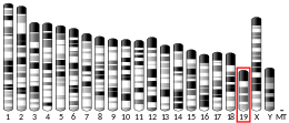
베스트로핀은 구조적으로 보존된 단백질의 고대 과로 박테리아에서 인간에 이르기까지 거의 모든 유기체에서 확인되었다.인간의 경우 칼슘 활성화 음이온 채널로 기능하며, 음이온 채널은 각각 몸 전체에 걸쳐 독특한 조직 분포를 가지고 있습니다.구체적으로, 11q13 염색체의 BEST1 유전자는 [7]망막에서 발현이 가장 높은 인간의 BESTrophin-1 단백질을 암호화한다.
구조.
진
베스트로핀 유전자는 보존된 유전자 구조를 공유하며, 크기가 거의 동일한 8개의 RFP-TM 도메인 인코딩 엑손과 고도로 보존된 엑손 인트론 경계를 가지고 있습니다.4개의 베스트로핀 유전자는 각각 가변 [5]길이의 독특한 3-프라임 말단을 가지고 있다.
BEST1은 두 가지 독립적인 연구에서 마이크로프탈리아 관련 전사 [8][9]인자에 의해 조절되는 것으로 나타났다.
단백질
베스트로핀-1은 주로 눈의 [10]망막색소상피(RPE)에서 발견되는 일체형 막 단백질이다.RPE층 내에서는 주로 기저측 혈장막에 위치한다.단백질 결정화 구조는 이 단백질의 1차 이온 채널 기능과 칼슘 조절 [10][7]능력을 나타냅니다.베스트로핀-1은 585개의 아미노산으로 구성되며, N- 및 C-termini는 모두 세포 내에 위치한다.
Best1의 구조는 5개의 동일한 서브유닛으로 구성되어 있으며, 각 서브유닛은 4회 막에 걸쳐 있으며, 불변 아르그-페프로(RFP)[7][11][12] 모티브를 포함하여 방향족 잔류물이 다량 함유된 두 번째 막 통과 도메인을 통해 연속적인 깔때기 모양의 모공을 형성합니다.모공에는 다양한 무극성 소수성 아미노산이 줄지어 있습니다.모공의 구조와 구성 모두 작은 음이온만 채널을 통해 완전히 이동할 수 있도록 합니다.채널은 2개의 깔때기 역할을 합니다.음이온을 위한 반선택적이고 좁은 입구에서 시작하여, 양전하를 띤 더 큰 영역으로 열리고, 그 후 모공을 통과하는 음이온의 크기를 더욱 제한하는 더 좁은 경로로 이어집니다.칼슘 걸쇠는 채널의 큰 중간 부분 주위에 벨팅 메커니즘으로 작용합니다.칼슘 이온은 마지막 막 통과 [7][12]도메인 직후의 C 말단에서 칼슘 결합에 의한 배좌 변화에 의해 채널의 개폐를 제어한다.
조직 및 세포하 분포
BEST1 유전자의 발현 위치는 단백질 기능을 위해 필수적이며 잘못된 국소화는 종종 다양한 망막 퇴행성 질환과 관련이 있다.BEST1 유전자는 주로 망막 색소 상피의 세포질에서 Best1 단백질을 발현합니다.단백질은 전형적으로 세포막 근처의 소포에 포함되어 있다.또한 Best1 단백질이 소포체(단백질 및 지질 합성에 관여하는 세포 내 소기관)에서 국소화 및 생산된다는 것을 뒷받침하는 연구도 있다.Best1은 전형적으로 칼레티쿨린, 칼넥신 및 Stim-1과 같은 소포체에서 합성된 다른 단백질과 함께 발현된다.염화물 이온의 역수송에 칼슘 이온이 관여하는 것도 Best1이 세포 [10]내에 칼슘 저장소를 형성하는 데 관여한다는 생각을 뒷받침한다.
기능.
Best1은 주로 전압 [6][10][12]의존성이 없는 세포막에서 세포 내 칼슘 활성 염화물 채널로 기능합니다.최근 Best1은 음이온 채널로 동작하는 것으로 나타났습니다.
질병.
베스트 비텔형 황반디스트로피(BVMD)
Best의 황반 디스트로피(BVMD)는 가장 일반적인 Best1 관련 질환 중 하나입니다.BVMD는 일반적으로 어린이에게서 두드러지며 [6][10]눈에 리포푸신(지질 잔차) 병변이 축적되는 것으로 나타난다.진단은 일반적으로 망막 색소 상피의 기저외막에서 칼슘 채널의 활성화가 감소하는 비정상적인 전기 적층도를 따른다.BEST1 유전자의 돌연변이는 채널 기능의 상실로 이어지고 결국 망막 변성으로 [10]이어진다.비록 BVMD가 황반디스트로피의 상염색체 우성 형태이기는 하지만, 영향을 받은 가족들의 압도적 다수가 북유럽 [7][10]혈통 출신이지만, 영향을 받은 가족 내 및 가족 간에 표현력은 다양하다.일반적으로 이 증상을 가진 사람들은 5단계씩 악화되는 단계를 경험하지만 시기와 심각도는 크게 다릅니다.BVMD는 종종 단일 미센스 돌연변이에 의해 발생하지만 아미노산 결손도 확인되었다.[7]Best1 염화물 채널의 기능 상실은 BVMD와 관련된 가장 일반적인 문제들 중 몇 가지를 설명할 수 있습니다. 즉, 세포 내 이온 농도를 조절하고 전체 [13]세포량을 조절할 수 없는 것입니다.지금까지 100개 이상의 질병을 일으키는 돌연변이가 BVMD와 다른 많은 퇴행성 망막 [12]질환과 관련이 있다.
성충성 황반디스트로피(AVMD)
성인 비텔형 황반 디스트로피(AVMD)는 망막의 BVMD와 유사한 병변으로 구성된다.그러나 원인은 BVMD만큼 명확하지 않습니다.유전자 검사를 통해 AVMD를 진단할 수 없기 때문에 AVMD와 패턴디스트로피를 구별하기가 어렵다.또, BEST1 돌연변이에 의한 AVMD와 PRPH2 돌연변이에 의한 AVMD의 임상적 차이가 있는지도 불명확하다.AVMD는 보통 BVMD보다 시력 감퇴가 적으며 일반적으로 가족 [7]내에서는 발병하지 않는다.
상염색체 열성베스트로핀증(ARB)
상염색체 열성베스트로핀증(ARB)은 2008년에 처음 확인되었다.ARB에 걸린 사람들은 생후 10년 동안 시력 저하를 보인다.부모와 가족은 일반적으로 질병이 상염색체 열성이기 때문에 BEST1 유전자의 양쪽 대립 유전자가 돌연변이가 되어야 함을 나타낸다.비텔형 병변은 종종 존재하며 어떤 경우에는 낭포성 황반부종을 수반한다.또한 다른 합병증도 관찰되었습니다.시력은 시간이 지남에 따라 서서히 감소하지만 감소 속도는 다양합니다.ARB를 일으키는 돌연변이는 비코딩 [7]영역에서 미센스 돌연변이를 일으키는 단일 염기 돌연변이에 이르기까지 다양합니다.
상염색체우성 유리정맥증
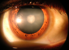
상염색체 우성 유리포상증은 1982년에 처음 확인되었으며 과도한 액체와 눈의 망막 색소 침착 변화로 인해 말초 시력이 저하되어 양쪽 눈에 나타난다.조기 발병성 백내장 또한 가능성이 [7]높다.
색소성 망막염(RP)
망막색소증은 2009년에 BEST1 유전자와 관련하여 처음 기술되었고 사람들의 BEST1 유전자의 4가지 다른 미센스 돌연변이와 관련이 있는 것으로 밝혀졌다.영향을 받는 모든 사람은 망막 내의 빛에 대한 반응 감소를 경험하고 색소 침착, 광시계 디스크, 체액 축적 [7]및 시력 저하가 있을 수 있다.
위의 모든 질병에는 알려진 치료법이나 치료법이 없습니다.그러나 2017년 현재, 연구자들은 망막 색소 [7]상피의 줄기세포 이식을 통한 치료법을 발견하는 작업을 진행 중이다.
레퍼런스
- ^ a b c GRCh38: 앙상블 릴리즈 89: ENSG00000167995 - 앙상블, 2017년 5월
- ^ a b c GRCm38: 앙상블 릴리즈 89: ENSMUSG000037418 - 앙상블, 2017년 5월
- ^ "Human PubMed Reference:". National Center for Biotechnology Information, U.S. National Library of Medicine.
- ^ "Mouse PubMed Reference:". National Center for Biotechnology Information, U.S. National Library of Medicine.
- ^ a b "Entrez Gene: BEST1 bestrophin 1".
- ^ a b c Kunzelmann K (September 2015). "TMEM16, LRRC8A, bestrophin: chloride channels controlled by Ca(2+) and cell volume". review. Trends in Biochemical Sciences. 40 (9): 535–43. doi:10.1016/j.tibs.2015.07.005. PMID 26254230.
- ^ a b c d e f g h i j k l m Johnson AA, Guziewicz KE, Lee CJ, Kalathur RC, Pulido JS, Marmorstein LY, Marmorstein AD (January 2017). "Bestrophin 1 and retinal disease". review. Progress in Retinal and Eye Research. 58: 45–69. doi:10.1016/j.preteyeres.2017.01.006. PMC 5600499. PMID 28153808.
- ^ Esumi N, Kachi S, Campochiaro PA, Zack DJ (January 2007). "VMD2 promoter requires two proximal E-box sites for its activity in vivo and is regulated by the MITF-TFE family". primary. The Journal of Biological Chemistry. 282 (3): 1838–50. doi:10.1074/jbc.M609517200. PMID 17085443.
- ^ Hoek KS, Schlegel NC, Eichhoff OM, Widmer DS, Praetorius C, Einarsson SO, Valgeirsdottir S, Bergsteinsdottir K, Schepsky A, Dummer R, Steingrimsson E (December 2008). "Novel MITF targets identified using a two-step DNA microarray strategy". primary. Pigment Cell & Melanoma Research. 21 (6): 665–76. doi:10.1111/j.1755-148X.2008.00505.x. PMID 19067971. S2CID 24698373.
- ^ a b c d e f g Strauss O, Neussert R, Müller C, Milenkovic VM (2012). "A potential cytosolic function of bestrophin-1". Retinal Degenerative Diseases. review. Advances in Experimental Medicine and Biology. Vol. 723. pp. 603–10. doi:10.1007/978-1-4614-0631-0_77. ISBN 978-1-4614-0630-3. PMID 22183384.
- ^ Hartzell HC, Qu Z, Yu K, Xiao Q, Chien LT (April 2008). "Molecular physiology of bestrophins: multifunctional membrane proteins linked to best disease and other retinopathies". review. Physiological Reviews. 88 (2): 639–72. doi:10.1152/physrev.00022.2007. PMID 18391176.
- ^ a b c d Xiao Q, Hartzell HC, Yu K (July 2010). "Bestrophins and retinopathies". review. Pflügers Archiv. 460 (2): 559–69. doi:10.1007/s00424-010-0821-5. PMC 2893225. PMID 20349192.
- ^ Olaf S, Müller C, Reichhart N, Tamm ER, Gomez NM (2014). "The Role of Bestrophin-1 in Intracellular Ca2+ Signaling". In Ash J, Grimm C, Hollyfield JG, Anderson RE, LaVail NM, Rickman CB (eds.). Retinal Degenerative Diseases: Mechanisms and Experimental Therapy. review. Advances in Experimental Medicine and Biology. Vol. 801. New York: Springer. pp. 113–119. doi:10.1007/978-1-4614-3209-8_15. ISBN 978-1-4614-3209-8. PMID 24664688.
추가 정보
- White K, Marquardt A, Weber BH (2000). "VMD2 mutations in vitelliform macular dystrophy (Best disease) and other maculopathies". Human Mutation. 15 (4): 301–8. doi:10.1002/(SICI)1098-1004(200004)15:4<301::AID-HUMU1>3.0.CO;2-N. PMID 10737974.
- Nordström S, Barkman Y (February 1977). "Hereditary maculardegeneration (HMD) in 246 cases traced to one gene-source in central Sweden". Hereditas. 84 (2): 163–76. doi:10.1111/j.1601-5223.1977.tb01394.x. PMID 838599.
- Forsman K, Graff C, Nordström S, Johansson K, Westermark E, Lundgren E, Gustavson KH, Wadelius C, Holmgren G (September 1992). "The gene for Best's macular dystrophy is located at 11q13 in a Swedish family". Clinical Genetics. 42 (3): 156–9. doi:10.1111/j.1399-0004.1992.tb03229.x. PMID 1395087. S2CID 924428.
- Stöhr H, Marquardt A, Rivera A, Cooper PR, Nowak NJ, Shows TB, Gerhard DS, Weber BH (January 1998). "A gene map of the Best's vitelliform macular dystrophy region in chromosome 11q12-q13.1". Genome Research. 8 (1): 48–56. doi:10.1101/gr.8.1.48. PMC 310689. PMID 9445487.
- Petrukhin K, Koisti MJ, Bakall B, Li W, Xie G, Marknell T, Sandgren O, Forsman K, Holmgren G, Andreasson S, Vujic M, Bergen AA, McGarty-Dugan V, Figueroa D, Austin CP, Metzker ML, Caskey CT, Wadelius C (July 1998). "Identification of the gene responsible for Best macular dystrophy". Nature Genetics. 19 (3): 241–7. doi:10.1038/915. PMID 9662395. S2CID 23334668.
- Pennisi E (July 1998). "New gene found for inherited macular degeneration". Science. 281 (5373): 31. doi:10.1126/science.281.5373.31. PMID 9679014. S2CID 44271444.
- Marquardt A, Stöhr H, Passmore LA, Krämer F, Rivera A, Weber BH (September 1998). "Mutations in a novel gene, VMD2, encoding a protein of unknown properties cause juvenile-onset vitelliform macular dystrophy (Best's disease)". Human Molecular Genetics. 7 (9): 1517–25. doi:10.1093/hmg/7.9.1517. PMID 9700209.
- Caldwell GM, Kakuk LE, Griesinger IB, Simpson SA, Nowak NJ, Small KW, Maumenee IH, Rosenfeld PJ, Sieving PA, Shows TB, Ayyagari R (May 1999). "Bestrophin gene mutations in patients with Best vitelliform macular dystrophy". Genomics. 58 (1): 98–101. doi:10.1006/geno.1999.5808. PMID 10331951.
- Bakall B, Marknell T, Ingvast S, Koisti MJ, Sandgren O, Li W, Bergen AA, Andreasson S, Rosenberg T, Petrukhin K, Wadelius C (May 1999). "The mutation spectrum of the bestrophin protein--functional implications". Human Genetics. 104 (5): 383–9. doi:10.1007/s004390050972. PMID 10394929. S2CID 21255716.
- Allikmets R, Seddon JM, Bernstein PS, Hutchinson A, Atkinson A, Sharma S, Gerrard B, Li W, Metzker ML, Wadelius C, Caskey CT, Dean M, Petrukhin K (June 1999). "Evaluation of the Best disease gene in patients with age-related macular degeneration and other maculopathies". Human Genetics. 104 (6): 449–53. doi:10.1007/s004390050986. PMID 10453731. S2CID 6214287.
- Palomba G, Rozzo C, Angius A, Pierrottet CO, Orzalesi N, Pirastu M (February 2000). "A novel spontaneous missense mutation in VMD2 gene is a cause of a best macular dystrophy sporadic case". American Journal of Ophthalmology. 129 (2): 260–2. doi:10.1016/S0002-9394(99)00327-X. PMID 10682987.
- Lotery AJ, Namperumalsamy P, Jacobson SG, Weleber RG, Fishman GA, Musarella MA, Hoyt CS, Héon E, Levin A, Jan J, Lam B, Carr RE, Franklin A, Radha S, Andorf JL, Sheffield VC, Stone EM (April 2000). "Mutation analysis of 3 genes in patients with Leber congenital amaurosis". Archives of Ophthalmology. 118 (4): 538–43. doi:10.1001/archopht.118.4.538. PMID 10766140.
- Lotery AJ, Munier FL, Fishman GA, Weleber RG, Jacobson SG, Affatigato LM, Nichols BE, Schorderet DF, Sheffield VC, Stone EM (May 2000). "Allelic variation in the VMD2 gene in best disease and age-related macular degeneration". Investigative Ophthalmology & Visual Science. 41 (6): 1291–6. PMID 10798642.
- Krämer F, White K, Pauleikhoff D, Gehrig A, Passmore L, Rivera A, Rudolph G, Kellner U, Andrassi M, Lorenz B, Rohrschneider K, Blankenagel A, Jurklies B, Schilling H, Schütt F, Holz FG, Weber BH (April 2000). "Mutations in the VMD2 gene are associated with juvenile-onset vitelliform macular dystrophy (Best disease) and adult vitelliform macular dystrophy but not age-related macular degeneration". European Journal of Human Genetics. 8 (4): 286–92. doi:10.1038/sj.ejhg.5200447. PMID 10854112.
- Marmorstein AD, Marmorstein LY, Rayborn M, Wang X, Hollyfield JG, Petrukhin K (November 2000). "Bestrophin, the product of the Best vitelliform macular dystrophy gene (VMD2), localizes to the basolateral plasma membrane of the retinal pigment epithelium". Proceedings of the National Academy of Sciences of the United States of America. 97 (23): 12758–63. Bibcode:2000PNAS...9712758M. doi:10.1073/pnas.220402097. PMC 18837. PMID 11050159.
- Marchant D, Gogat K, Boutboul S, Péquignot M, Sternberg C, Dureau P, Roche O, Uteza Y, Hache JC, Puech B, Puech V, Dumur V, Mouillon M, Munier FL, Schorderet DF, Marsac C, Dufier JL, Abitbol M (March 2001). "Identification of novel VMD2 gene mutations in patients with best vitelliform macular dystrophy". Human Mutation. 17 (3): 235. doi:10.1002/humu.9. PMID 11241846. S2CID 16128731.
- Eksandh L, Bakall B, Bauer B, Wadelius C, Andréasson S (June 2001). "Best's vitelliform macular dystrophy caused by a new mutation (Val89Ala) in the VMD2 gene". Ophthalmic Genetics. 22 (2): 107–15. doi:10.1076/opge.22.2.107.2226. PMID 11449320. S2CID 7035792.
- Sun H, Tsunenari T, Yau KW, Nathans J (March 2002). "The vitelliform macular dystrophy protein defines a new family of chloride channels". Proceedings of the National Academy of Sciences of the United States of America. 99 (6): 4008–13. Bibcode:2002PNAS...99.4008S. doi:10.1073/pnas.052692999. PMC 122639. PMID 11904445.
- Xiao Q, Yu K, Cui YY, Hartzell HC (September 2009). "Dysregulation of human bestrophin-1 by ceramide-induced dephosphorylation". The Journal of Physiology. 587 (Pt 18): 4379–91. doi:10.1113/jphysiol.2009.176800. PMC 2766645. PMID 19635817.
- Xiao Q, Prussia A, Yu K, Cui YY, Hartzell HC (December 2008). "Regulation of bestrophin Cl channels by calcium: role of the C terminus". The Journal of General Physiology. 132 (6): 681–92. doi:10.1085/jgp.200810056. PMC 2585866. PMID 19029375.
외부 링크
- 색소성 망막염에 대한 Gen Reviews/NCBI/NIH/UW 엔트리 개요
- UCSC Genome Browser의 Human BEST1 게놈 위치 및 BEST1 유전자 상세 페이지.


